6K5J image
Deposition Date 2019-05-29
Release Date 2019-09-25
Last Version Date 2024-10-23
Entry Detail
PDB ID:
6K5J
Keywords:
Title:
Structure of a glycoside hydrolase family 3 beta-N-acetylglucosaminidase from Paenibacillus sp. str. FPU-7
Biological Source:
Source Organism:
Host Organism:
Method Details:
Experimental Method:
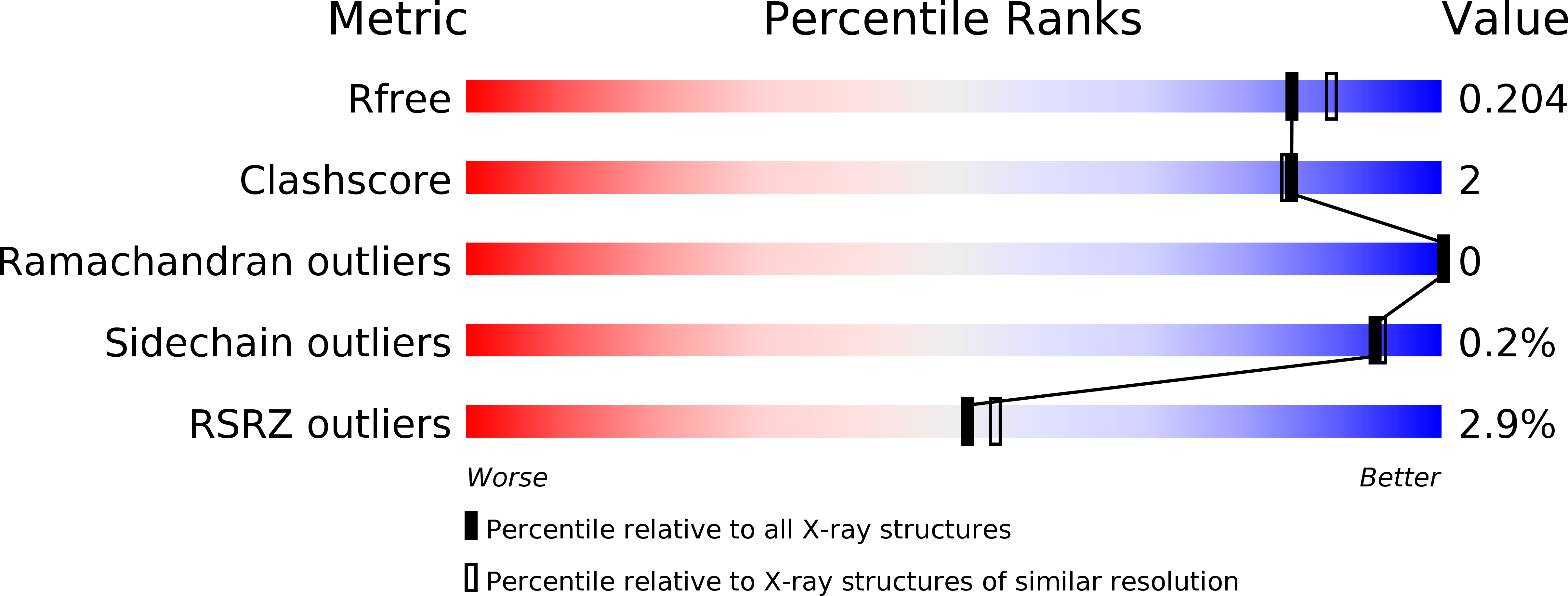
Resolution:
1.90 Å
R-Value Free:
0.20
R-Value Work:
0.16
R-Value Observed:
0.16
Space Group:
P 43 21 2
Macromolecular Entities
Polymer Type:polypeptide(L)
Molecule:GH3 beta-N-acetylglucosaminidase
Chain IDs:A
Chain Length:535
Number of Molecules:1
Biological Source:Paenibacillaceae
Primary Citation
Structural and functional characterization of a glycoside hydrolase family 3 beta-N-acetylglucosaminidase from Paenibacillus sp. str. FPU-7.
J.Biochem. 166 503 515 (2019)
PMID: 31501879 DOI: 10.1093/jb/mvz072

Abstact

Chitin, a β-1,4-linked homopolysaccharide of N-acetyl-d-glucosamine (GlcNAc), is one of the most abundant biopolymers on Earth. Paenibacillus sp. str. FPU-7 produces several different chitinases and converts chitin into N,N'-diacetylchitobiose ((GlcNAc)2) in the culture medium. However, the mechanism by which the Paenibacillus species imports (GlcNAc)2 into the cytoplasm and divides it into the monomer GlcNAc remains unclear. The gene encoding Paenibacillus β-N-acetyl-d-glucosaminidase (PsNagA) was identified in the Paenibacillus sp. str. FPU-7 genome using an expression cloning system. The deduced amino acid sequence of PsNagA suggests that the enzyme is a part of the glycoside hydrolase family 3 (GH3). Recombinant PsNagA was successfully overexpressed in Escherichia coli and purified to homogeneity. As assessed by gel permeation chromatography, the enzyme exists as a 57-kDa monomer. PsNagA specifically hydrolyses chitin oligosaccharides, (GlcNAc)2-4, 4-nitrophenyl N-acetyl β-d-glucosamine (pNP-GlcNAc) and pNP-(GlcNAc)2-6, but has no detectable activity against 4-nitrophenyl β-d-glucose, 4-nitrophenyl β-d-galactosamine and colloidal chitin. In this study, we present a 1.9 Å crystal structure of PsNagA bound to GlcNAc. The crystal structure reveals structural features related to substrate recognition and the catalytic mechanism of PsNagA. This is the first study on the structural and functional characterization of a GH3 β-N-acetyl-d-glucosaminidase from Paenibacillus sp.

Legend

Protein

Chemical

Disease

Primary Citation of related structures