6K3G image
Deposition Date 2019-05-18
Release Date 2020-03-25
Last Version Date 2023-11-22
Entry Detail
PDB ID:
6K3G
Keywords:
Title:
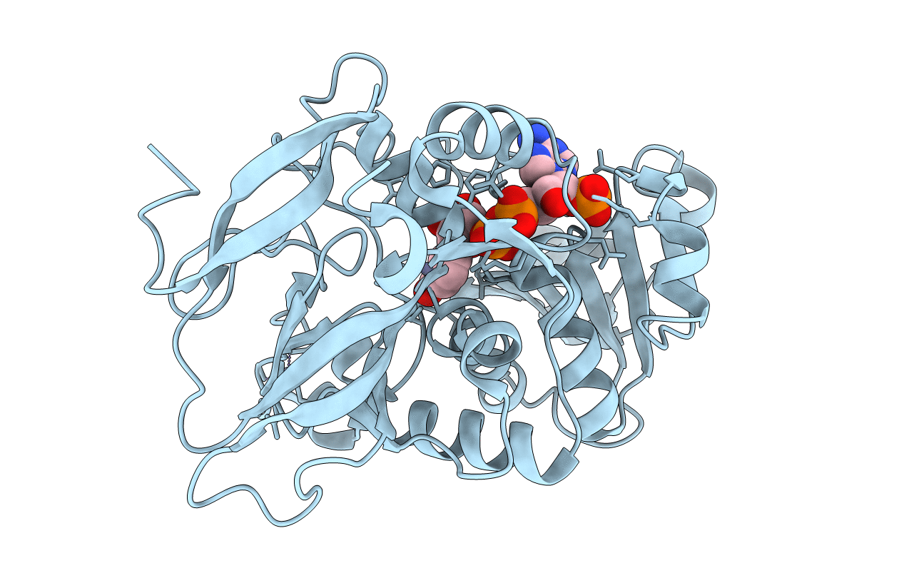
Crystal structure of 10-Hydroxygeraniol Dehydrogenase from Cantharanthus roseus in complex with NADP+
Biological Source:
Source Organism:
Host Organism:
Method Details:
Experimental Method:
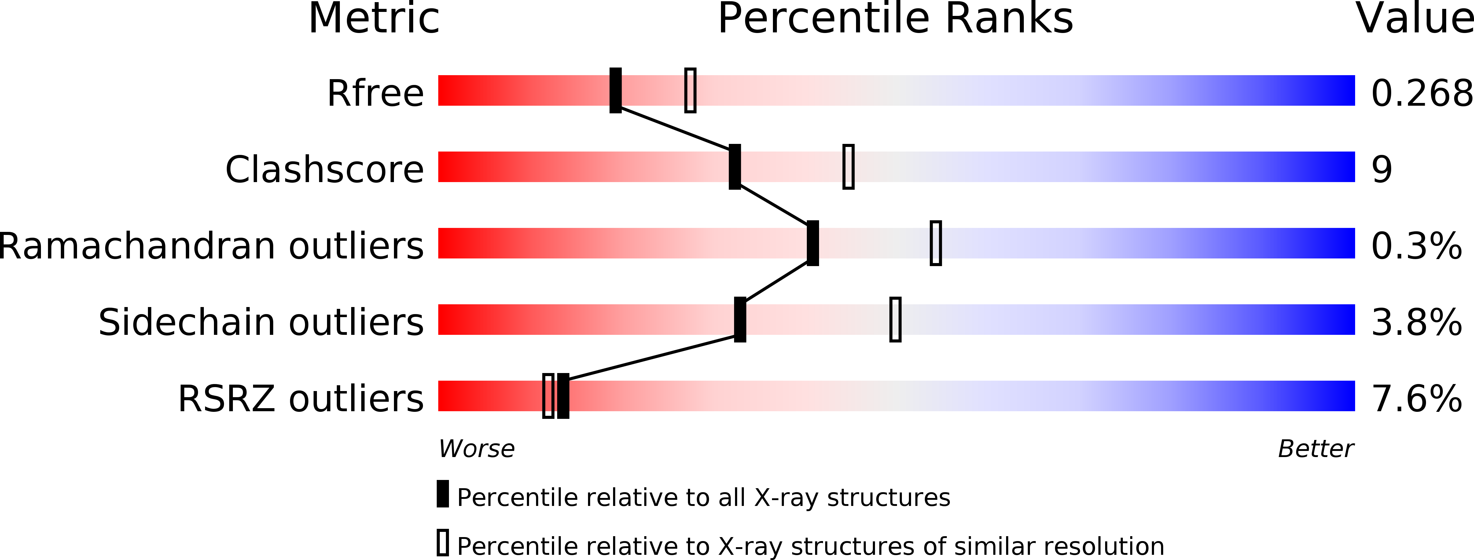
Resolution:
2.41 Å
R-Value Free:
0.26
R-Value Work:
0.20
R-Value Observed:
0.21
Space Group:
P 32 1 2
Macromolecular Entities
Structures with similar UniProt ID
Protein Blast
Polymer Type:polypeptide(L)
Molecule:10-hydroxygeraniol oxidoreductase
Chain IDs:A (auth: B)
Chain Length:360
Number of Molecules:1
Biological Source:Catharanthus roseus
Primary Citation
Structural studies on 10-hydroxygeraniol dehydrogenase: A novel linear substrate-specific dehydrogenase from Catharanthus roseus.
Proteins 88 1197 1206 (2020)
PMID: 32181958 DOI: 10.1002/prot.25891

Abstact

Conversion of 10-hydroxygeraniol to 10-oxogeranial is a crucial step in iridoid biosynthesis. This reaction is catalyzed by a zinc-dependent alcohol dehydrogenase, 10-hydroxygeraniol dehydrogenase, belonging to the family of medium-chain dehydrogenase/reductase (MDR). Here, we report the crystal structures of a novel 10-hydroxygeraniol dehydrogenase from Catharanthus roseus in its apo and nicotinamide adenine dinucleotide phosphate (NADP+) bound forms. Structural analysis and docking studies reveal how subtle conformational differences of loops L1, L2, L3, and helix α9' at the orifice of the catalytic site confer differential activity of the enzyme toward various substrates, by modulating the binding pocket shape and volume. The present study, first of its kind, provides insights into the structural basis of substrate specificity of MDRs specific to linear substrates. Furthermore, comparison of apo and NADP+ bound structures suggests that the enzyme adopts open and closed states to facilitate cofactor binding.

Legend

Protein

Chemical

Disease

Primary Citation of related structures
Feedback Form
Name
Email
Institute
Feedback