6K25 image
Deposition Date 2019-05-13
Release Date 2020-03-25
Last Version Date 2023-11-22
Entry Detail
PDB ID:
6K25
Title:
Crystal structure of Ca-unbound human Annexin A5 in low salt condition
Biological Source:
Source Organism:
Homo sapiens (Taxon ID: 9606)
Host Organism:
Method Details:
Experimental Method:
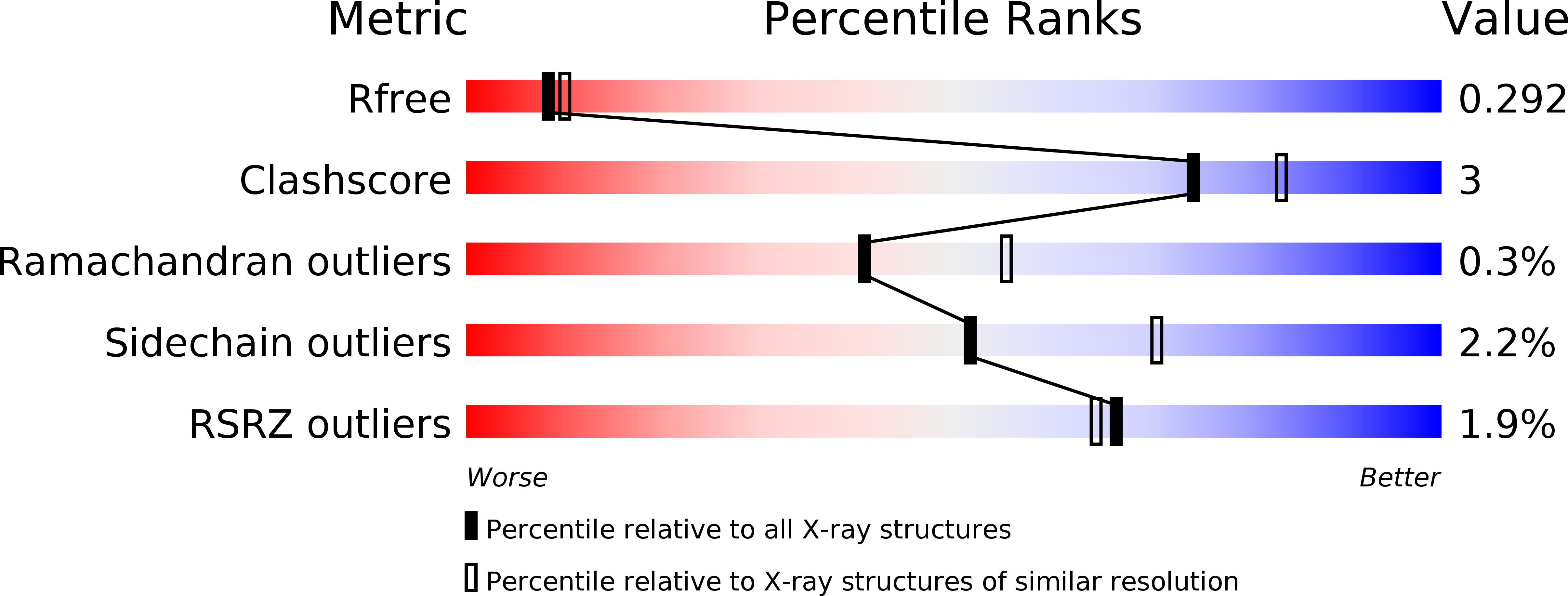
Resolution:
2.40 Å
R-Value Free:
0.29
R-Value Work:
0.22
R-Value Observed:
0.23
Space Group:
P 63 2 2
Macromolecular Entities
Structures with similar UniProt ID
Protein Blast
Polymer Type:polypeptide(L)
Molecule:Annexin A5
Gene (Uniprot):ANXA5
Chain IDs:A
Chain Length:331
Number of Molecules:1
Biological Source:Homo sapiens
Primary Citation
High-resolution structures of annexin A5 in a two-dimensional array.
J.Struct.Biol. 209 107401 107401 (2020)
PMID: 31605770 DOI: 10.1016/j.jsb.2019.10.003

Abstact

Annexins are soluble cytosolic proteins that bind to cell membranes. Annexin A5 self-assembles into a two-dimensional (2D) array and prevents cell rupture by attaching to damaged membranes. However, this process is not fully understood at the molecular level. In this study, we determined the crystal structures of annexin A5 with and without calcium (Ca2+) and confirmed the Ca2+-dependent outward motion of a tryptophan residue. Strikingly, the two structures exhibited the same crystal packing and 2D arrangement into a p3 lattice, which agrees well with the results of low-resolution structural imaging. High-resolution structures indicated that a three-fold interaction near the tryptophan residue is important for mediating the formation of the p3 lattice. A hypothesis on the promotion of p3 lattice formation by phosphatidyl serine (PS) is also suggested. This study provides molecular insight into how annexins modulate the physical properties of cell membranes as a function of Ca2+ concentration and the phospholipid composition of the membrane.

Legend

Protein

Chemical

Disease

Primary Citation of related structures