Deposition Date
2019-05-08
Release Date
2019-12-11
Last Version Date
2024-11-20
Entry Detail
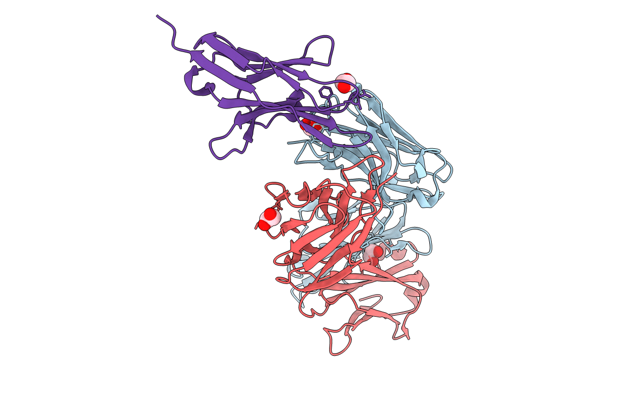
PDB ID:
6K0Y
Keywords:
Title:
Study of the interactions of a novel monoclonal antibody, mAb059c, with the hPD-1 receptor
Biological Source:
Source Organism(s):
Homo sapiens (Taxon ID: 9606)
Expression System(s):
Method Details:
Experimental Method:
Resolution:
1.70 Å
R-Value Free:
0.21
R-Value Work:
0.17
R-Value Observed:
0.17
Space Group:
P 21 21 21