6JX3 image
Deposition Date 2019-04-22
Release Date 2019-06-26
Last Version Date 2024-03-27
Entry Detail
PDB ID:
6JX3
Title:
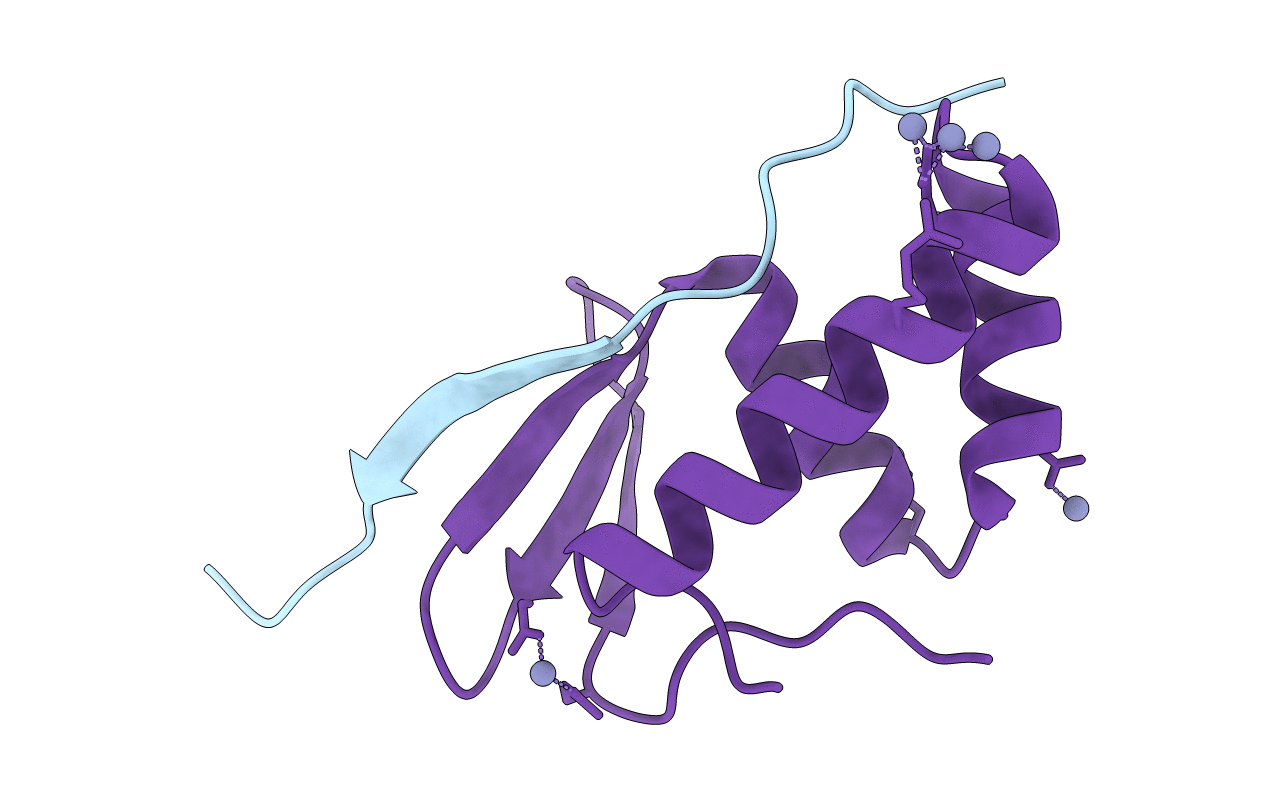
Lasso peptide synthetase B1 complexed with the leader peptide
Biological Source:
Source Organism:
Host Organism:
Method Details:
Experimental Method:
Resolution:
1.70 Å
R-Value Free:
0.20
R-Value Work:
0.18
R-Value Observed:
0.18
Space Group:
C 2 2 21
Macromolecular Entities
Polymer Type:polypeptide(L)
Molecule:TfuA-Leader
Chain IDs:B (auth: A)
Chain Length:22
Number of Molecules:1
Biological Source:Thermobifida fusca
Polymer Type:polypeptide(L)
Molecule:TfuB1
Gene (Uniprot):Tfu_1144
Chain IDs:A (auth: B)
Chain Length:99
Number of Molecules:1
Biological Source:Thermobifida fusca
Ligand Molecules
Primary Citation
Structural Basis of Leader Peptide Recognition in Lasso Peptide Biosynthesis Pathway.
Acs Chem.Biol. 14 1619 1627 (2019)
PMID: 31188556 DOI: 10.1021/acschembio.9b00348

Abstact

Lasso peptides are a class of ribosomally synthesized and post-translationally modified peptides (RiPPs) with a unique 3D-interlocked structure, in which an N-terminal macrolactam ring is threaded by a linear C-terminal part. The unique structure of lasso peptides is introduced into ribosomally translated precursor peptides by lasso peptide synthetase encompassing proteins B and C or B1, B2, and C when the B enzyme is split into two distinct proteins. The B1 protein recognizes the leader sequence of the precursor peptide, and then the B2 protein cleaves it. The C protein catalyzes the formation of the macrolactam ring. However, the detailed mechanism of lasso peptide maturation has remained elusive, due to the lack of structural information about the responsible proteins. Here we report the crystal structure of the B1 protein from the thermophilic actinobacteria, Thermobifida fusca (TfuB1), complexed with the leader peptide (TfuA-Leader), which revealed the detailed mechanism of leader peptide recognition. The structure of TfuB1 consists of an N-terminal β-sheet and three C-terminal helices. The leader peptide is docked on one edge of the N-terminal β-sheet of TfuB1, as an additional β strand. Three conserved amino acid residues of the leader peptide (TfuA Tyr-17, Pro-14, and Leu-12) fit well on the hydrophobic cleft between the β-sheet and adjacent helices. Biochemical analysis demonstrated that these conserved residues are essential for affinity between TfuB1 and the TfuA-Leader. Furthermore, we found that TfuB1 and the leader peptide jointly form a hydrophobic patch on the β-sheet, which includes the highly conserved TfuA Phe-6 and TfuB1 Tyr33. Homology modeling and mutational analysis of the B1 protein from a firmicute, Bacillus pseudomycoides (PsmB1), revealed that the hydrophobic patch is conserved in a wide range of species and involved in the cleavage activity of the B2 protein, indicating it forms the interaction surface for the B2 protein or the core part of the precursor peptide.

Legend

Protein

Chemical

Disease

Primary Citation of related structures