
Deposition Date
2019-04-15
Release Date
2020-03-25
Last Version Date
2023-11-22
Entry Detail
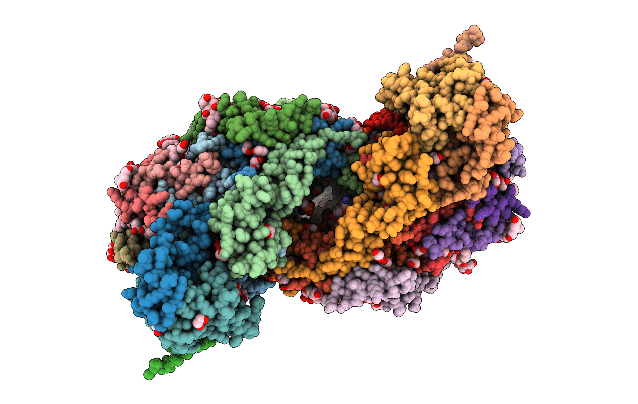
PDB ID:
6JUW
Keywords:
Title:
BOVINE HEART CYTOCHROME C OXIDASE IN CATALITIC INTERMEDIATES AT 1.80 ANGSTROM RESOLUTION
Biological Source:
Source Organism:
Bos taurus (Taxon ID: 9913)
Method Details:
Experimental Method:
Resolution:
1.80 Å
R-Value Free:
0.18
R-Value Work:
0.15
R-Value Observed:
0.15
Space Group:
P 21 21 21
