6JUI image
Deposition Date 2019-04-14
Release Date 2020-02-19
Last Version Date 2024-03-27
Entry Detail
PDB ID:
6JUI
Title:
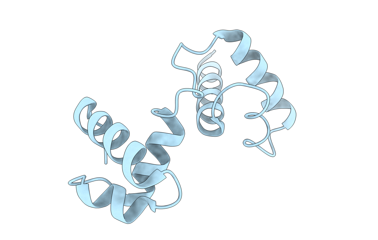
The atypical Myb-like protein Cdc5 contains two distinct nucleic acid-binding surfaces
Biological Source:
Source Organism:
Host Organism:
Method Details:
Experimental Method:
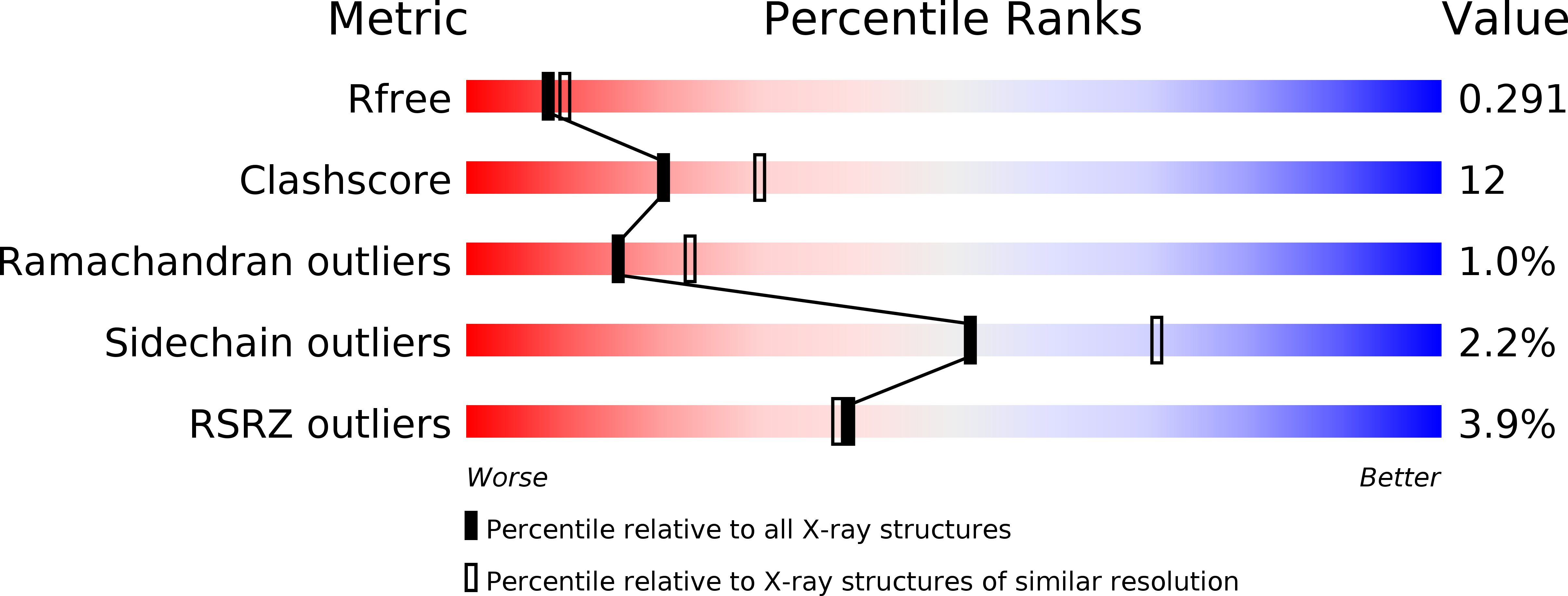
Resolution:
2.40 Å
R-Value Free:
0.27
R-Value Work:
0.23
R-Value Observed:
0.23
Space Group:
C 2 2 21
Macromolecular Entities
Structures with similar UniProt ID
Protein Blast
Polymer Type:polypeptide(L)
Molecule:Pre-mRNA-splicing factor CEF1
Gene (Uniprot):CEF1
Chain IDs:A
Chain Length:116
Number of Molecules:1
Biological Source:Magnaporthe oryzae
Primary Citation
Two distinct nucleic acid binding surfaces of Cdc5 regulate development.
Biochem.J. 476 3355 3368 (2019)
PMID: 31652438 DOI: 10.1042/BCJ20190502

Abstact

Cell division cycle 5 (Cdc5) is a highly conserved nucleic acid binding protein among eukaryotes and plays critical roles in development. Cdc5 can simultaneously bind to DNA and RNA by its N-terminal DNA-binding domain (DBD), but molecular mechanisms describing its nucleic acid recognition and the regulation of development through its nucleic acid binding remain unclear. Herein, we present a crystal structure of the N-terminal DBD of MoCdc5 (MoCdc5-DBD) from the rice blast fungus Magnaporthe oryzae. Residue K100 of MoCdc5 is on the periphery of a positively charged groove that is formed by K42, K45, R47, and N92 and is evolutionally conserved. Mutation of K100 significantly reduces the affinity of MoCdc5-DBD to a Cdc5-binding element but not to a conventional myeloblastosis (Myb) domain-binding element, suggesting that K100 is a key residue of the high binding affinity to Cdc5-binding element. Another conserved residue (R31) is located close to the U6 RNA in the structure of the spliceosome, and its mutation dramatically reduces the binding capacity of MoCdc5-DBD for U6 RNA. Importantly, mutations in these key residues, including R31, K42, and K100 in AtCDC5, an Arabidopsis thaliana ortholog of MoCdc5, greatly impair the functions of AtCDC5, resulting in pleiotropic development defects and reduced levels of primary microRNA transcripts. Taken together, our findings suggest that Cdc5-DBD binds nucleic acids with two distinct binding surfaces, one for DNA and another for RNA, which together contribute to establishing the regulation mechanism of Cdc5 on development through nucleic acid binding.

Legend

Protein

Chemical

Disease

Primary Citation of related structures