6JMV image
Deposition Date 2019-03-13
Release Date 2019-07-24
Last Version Date 2024-10-16
Entry Detail
PDB ID:
6JMV
Title:
Crystal structure of the GluK3 ligand binding domain complex with SYM and zinc
Biological Source:
Source Organism:
Host Organism:
Method Details:
Experimental Method:
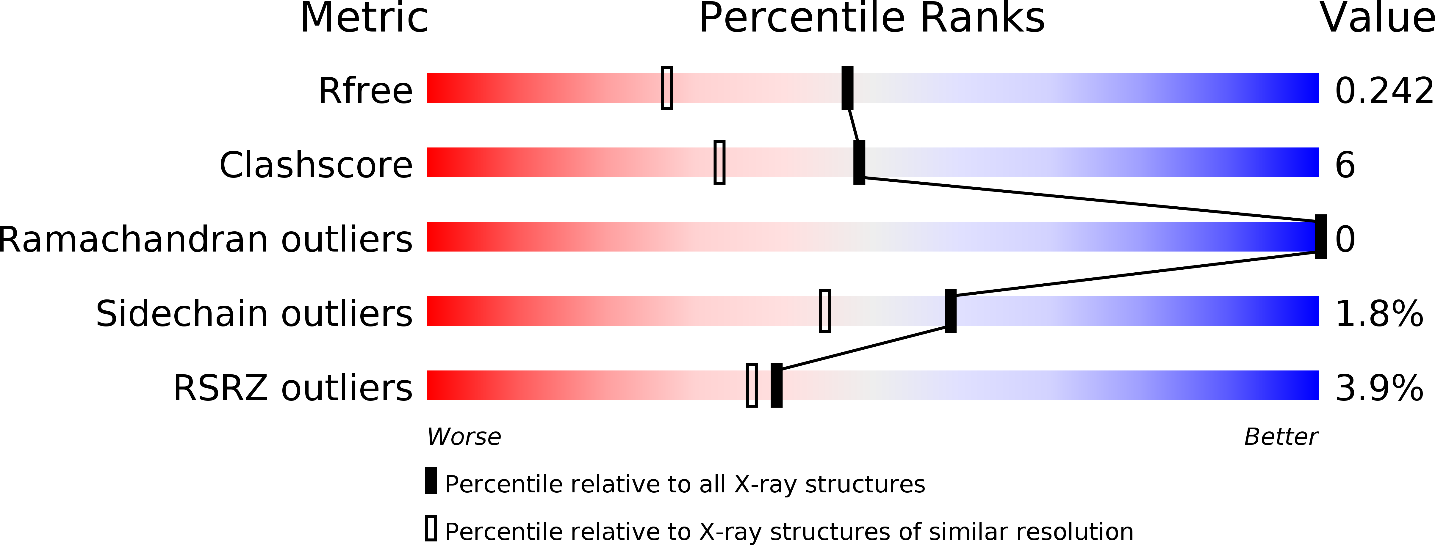
Resolution:
1.83 Å
R-Value Free:
0.24
R-Value Work:
0.20
R-Value Observed:
0.20
Space Group:
P 2 21 2
Macromolecular Entities
Polymer Type:polypeptide(L)
Molecule:Glutamate receptor ionotropic, kainate 3
Gene (Uniprot):Grik3
Chain IDs:A, B
Chain Length:257
Number of Molecules:2
Biological Source:Rattus norvegicus
Primary Citation
Structural and Functional Insights into GluK3-kainate Receptor Desensitization and Recovery.
Sci Rep 9 10254 10254 (2019)
PMID: 31311973 DOI: 10.1038/s41598-019-46770-z

Abstact

GluK3-kainate receptors are atypical members of the iGluR family that reside at both the pre- and postsynapse and play a vital role in the regulation of synaptic transmission. For a better understanding of structural changes that underlie receptor functions, GluK3 receptors were trapped in desensitized and resting/closed states and structures analyzed using single particle cryo-electron microscopy. While the desensitized GluK3 has domain organization as seen earlier for another kainate receptor-GluK2, antagonist bound GluK3 trapped a resting state with only two LBD domains in dimeric arrangement necessary for receptor activation. Using structures as a guide, we show that the N-linked glycans at the interface of GluK3 ATD and LBD likely mediate inter-domain interactions and attune receptor-gating properties. The mutational analysis also identified putative N-glycan interacting residues. Our results provide a molecular framework for understanding gating properties unique to GluK3 and exploring the role of N-linked glycosylation in their modulation.

Legend

Protein

Chemical

Disease

Primary Citation of related structures