Deposition Date
2019-03-05
Release Date
2019-07-10
Last Version Date
2024-10-16
Entry Detail
PDB ID:
6JLD
Keywords:
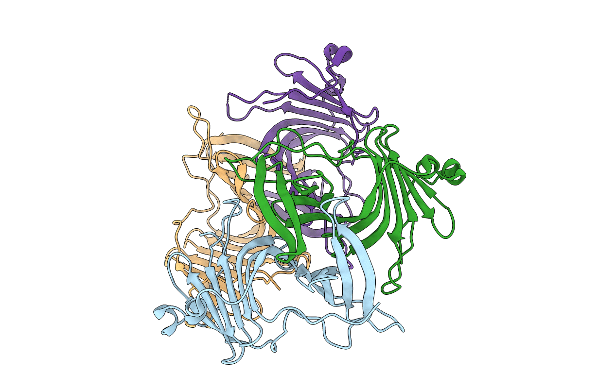
Title:
Crystal structure of a human ependymin related protein
Biological Source:
Source Organism(s):
Homo sapiens (Taxon ID: 9606)
Expression System(s):
Method Details:
Experimental Method:
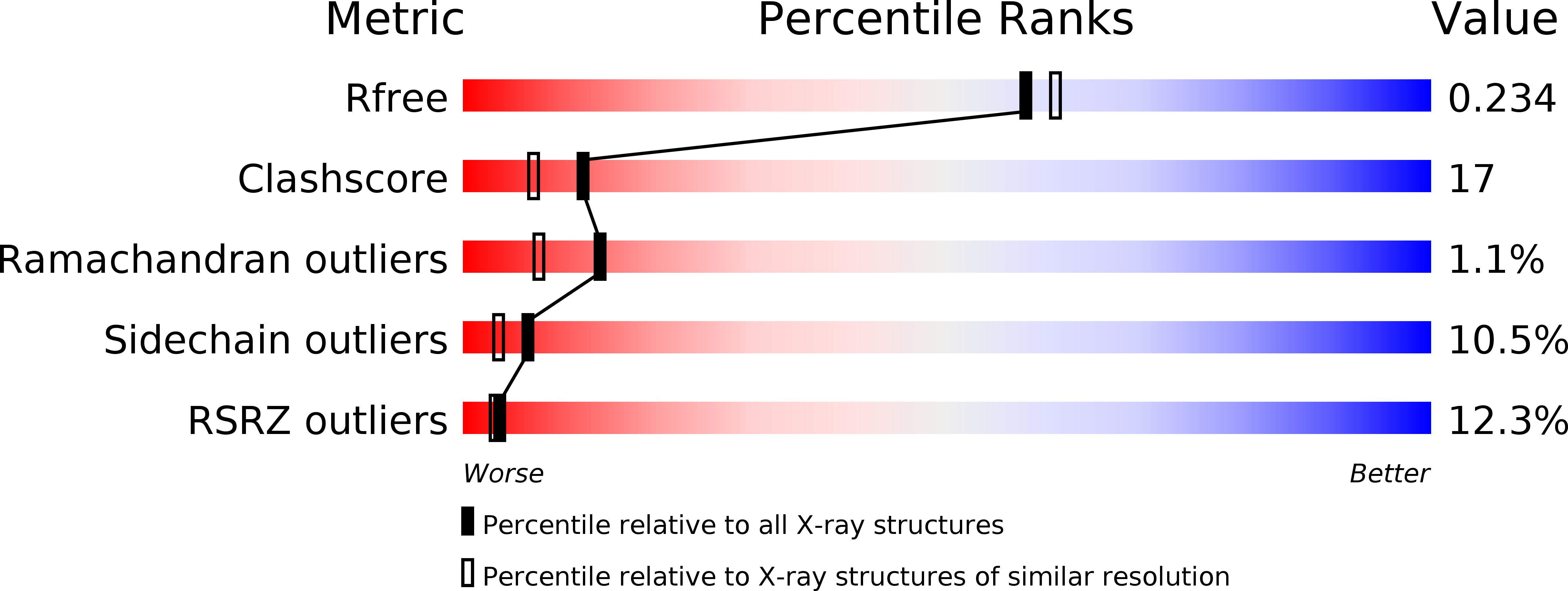
Resolution:
2.00 Å
R-Value Free:
0.23
R-Value Work:
0.19
R-Value Observed:
0.20
Space Group:
P 43