6JGJ image
Deposition Date 2019-02-14
Release Date 2019-04-17
Last Version Date 2024-10-23
Entry Detail
PDB ID:
6JGJ
Title:
Crystal structure of the F99S/M153T/V163A/E222Q variant of GFP at 0.78 A
Biological Source:
Source Organism:
Method Details:
Experimental Method:
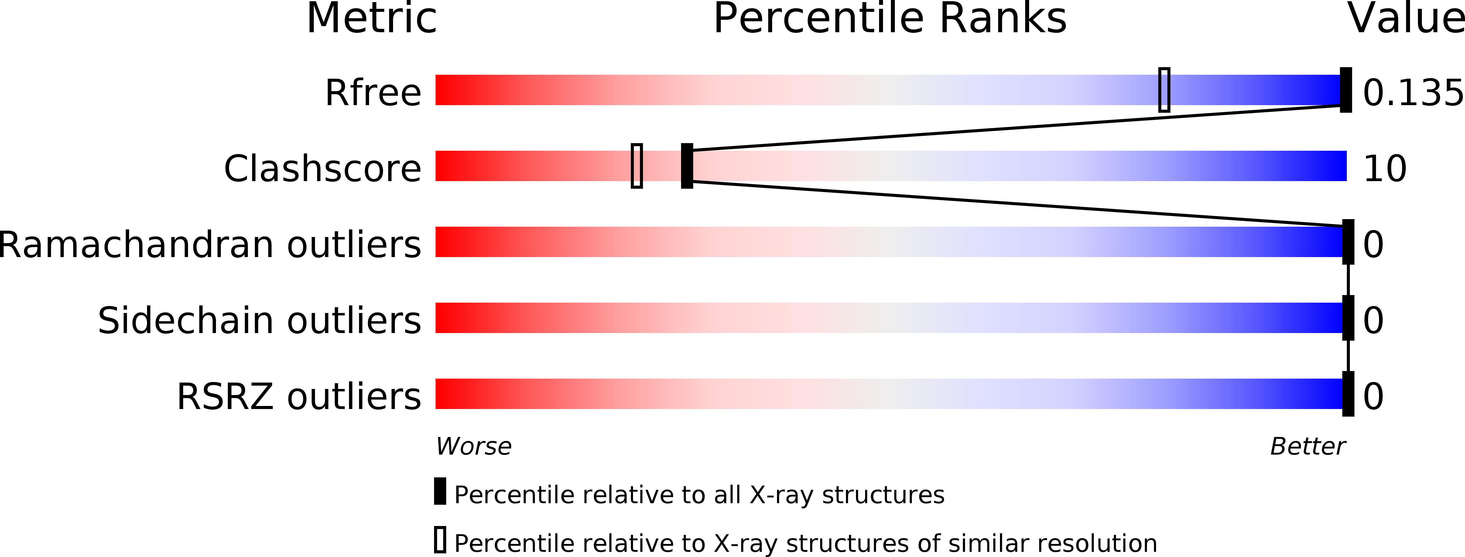
Resolution:
0.78 Å
R-Value Free:
0.12
R-Value Work:
0.10
Space Group:
P 21 21 21
Macromolecular Entities
Structures with similar UniProt ID
Protein Blast
Polymer Type:polypeptide(L)
Molecule:Green fluorescent protein
Gene (Uniprot):GFP
Mutagens:E222Q, F99S, M153T, V163A
Chain IDs:A
Chain Length:230
Number of Molecules:1
Biological Source:Aequorea victoria
Modified Residue
Compound ID Chain ID Parent Comp ID Details 2D Image
GYS A SER chromophore
Ligand Molecules
Primary Citation
Subatomic resolution X-ray structures of green fluorescent protein.
Iucrj 6 387 400 (2019)
PMID: 31098020 DOI: 10.1107/S205225251900246X

Abstact

Green fluorescent protein (GFP) is a light-emitting protein that does not require a prosthetic group for its fluorescent activity. As such, GFP has become indispensable as a molecular tool in molecular biology. Nonetheless, there has been no subatomic elucidation of the GFP structure owing to the structural polymorphism around the chromophore. Here, subatomic resolution X-ray structures of GFP without the structural polymorphism are reported. The positions of H atoms, hydrogen-bonding network patterns and accurate geometric parameters were determined for the two protonated forms. Compared with previously determined crystal structures and theoretically optimized structures, the anionic chromophores of the structures represent the authentic resonance state of GFP. In addition, charge-density analysis based on atoms-in-molecules theory and noncovalent interaction analysis highlight weak but substantial interactions between the chromophore and the protein environment. Considered with the derived chemical indicators, the lone pair-π interactions between the chromophore and Thr62 should play a sufficient role in maintaining the electronic state of the chromophore. These results not only reveal the fine structural features that are critical to understanding the properties of GFP, but also highlight the limitations of current quantum-chemical calculations.

Legend

Protein

Chemical

Disease

Primary Citation of related structures
Feedback Form
Name
Email
Institute
Feedback