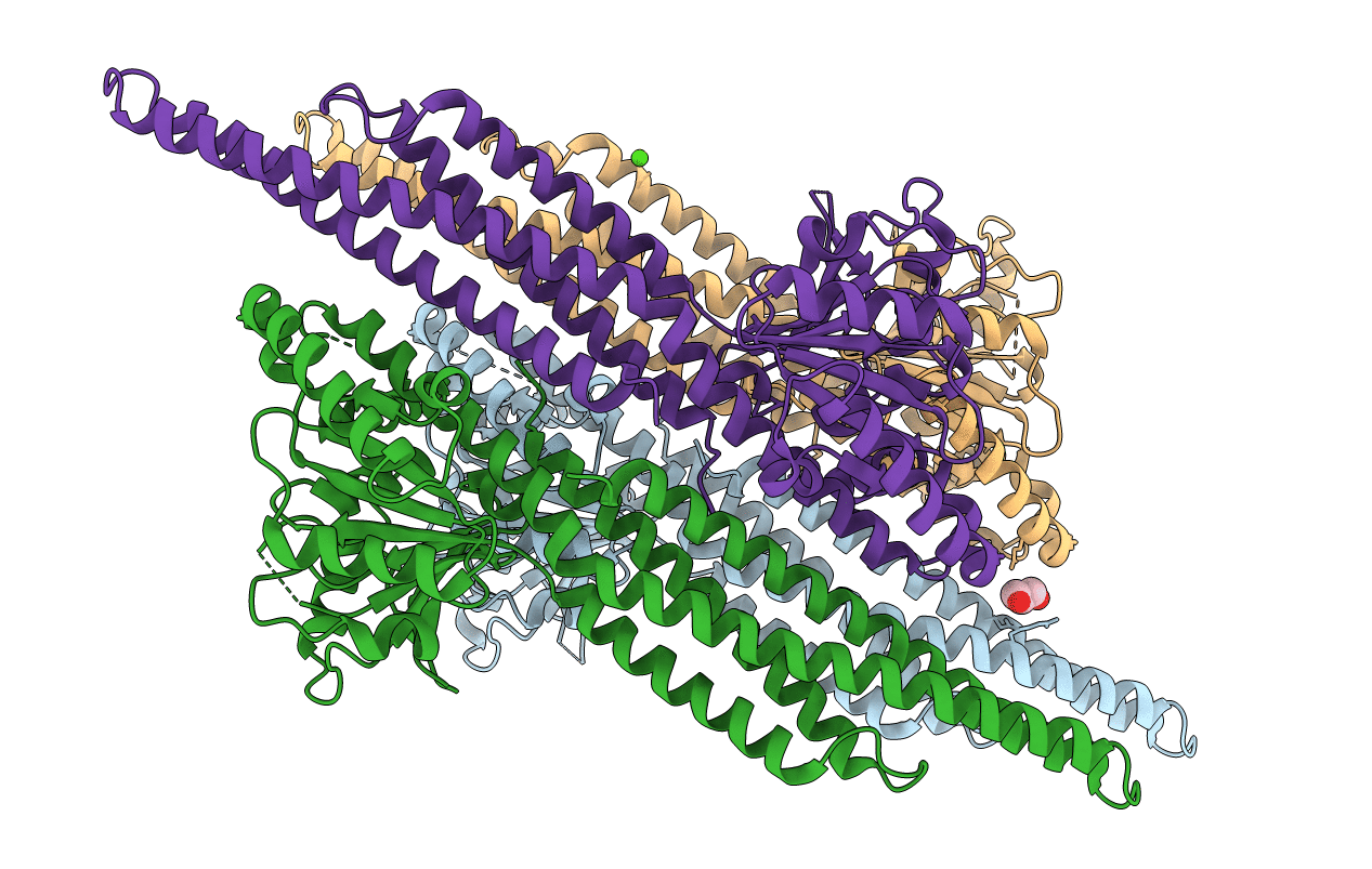
6JFL image
Deposition Date 2019-02-10
Release Date 2019-11-13
Last Version Date 2023-11-22
Entry Detail
PDB ID:
6JFL
Title:
Nucleotide-free Mitofusin2 (MFN2)
Biological Source:
Source Organism:
Homo sapiens (Taxon ID: 9606)
Host Organism:
Method Details:
Experimental Method:
Resolution:
2.81 Å
R-Value Free:
0.28
R-Value Work:
0.20
R-Value Observed:
0.21
Space Group:
P 1 21 1
Macromolecular Entities
Polymer Type:polypeptide(L)
Molecule:Mitofusin-2,cDNA FLJ57997, highly similar to Transmembrane GTPase MFN2
Gene (Uniprot):MFN2
Chain IDs:A, B, C, D
Chain Length:438
Number of Molecules:4
Biological Source:Homo sapiens
Primary Citation
Structural insights of human mitofusin-2 into mitochondrial fusion and CMT2A onset.
Nat Commun 10 4914 4914 (2019)
PMID: 31664033 DOI: 10.1038/s41467-019-12912-0

Abstact

Mitofusin-2 (MFN2) is a dynamin-like GTPase that plays a central role in regulating mitochondrial fusion and cell metabolism. Mutations in MFN2 cause the neurodegenerative disease Charcot-Marie-Tooth type 2A (CMT2A). The molecular basis underlying the physiological and pathological relevance of MFN2 is unclear. Here, we present crystal structures of truncated human MFN2 in different nucleotide-loading states. Unlike other dynamin superfamily members including MFN1, MFN2 forms sustained dimers even after GTP hydrolysis via the GTPase domain (G) interface, which accounts for its high membrane-tethering efficiency. The biochemical discrepancy between human MFN2 and MFN1 largely derives from a primate-only single amino acid variance. MFN2 and MFN1 can form heterodimers via the G interface in a nucleotide-dependent manner. CMT2A-related mutations, mapping to different functional zones of MFN2, lead to changes in GTP hydrolysis and homo/hetero-association ability. Our study provides fundamental insight into how mitofusins mediate mitochondrial fusion and the ways their disruptions cause disease.

Legend

Protein

Chemical

Disease

Primary Citation of related structures