6JDG image
Deposition Date 2019-02-01
Release Date 2019-12-11
Last Version Date 2023-11-22
Entry Detail
PDB ID:
6JDG
Title:
Complexed crystal structure of PaSSB with ssDNA dT20 at 2.39 angstrom resolution
Biological Source:
Source Organism:
Host Organism:
Method Details:
Experimental Method:
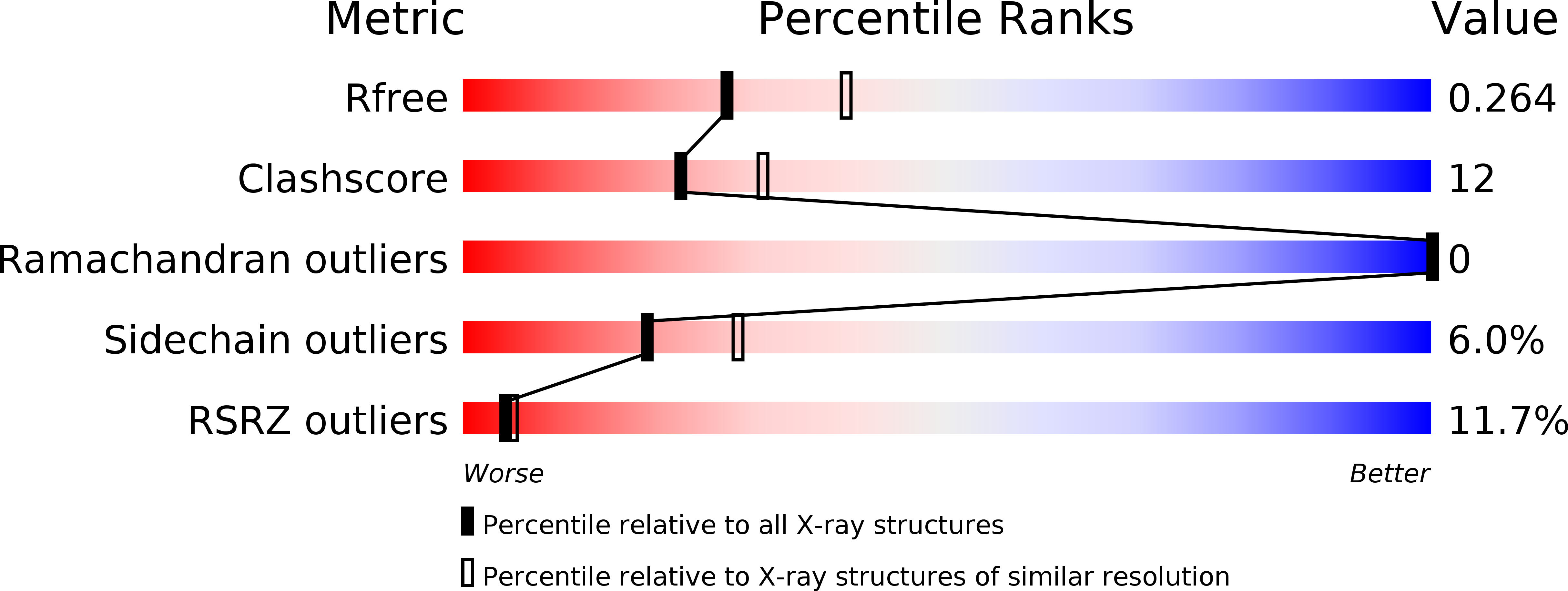
Resolution:
2.39 Å
R-Value Free:
0.26
R-Value Work:
0.21
R-Value Observed:
0.21
Space Group:
P 31
Macromolecular Entities
Polymer Type:polypeptide(L)
Molecule:Single-stranded DNA-binding protein
Gene (Uniprot):ssb
Chain IDs:A, B, C, D
Chain Length:121
Number of Molecules:4
Biological Source:Pseudomonas aeruginosa PAO1
Polymer Type:polydeoxyribonucleotide
Molecule:DNA (5'-D(P*TP*TP*TP*TP*TP*TP*TP*TP*TP*TP*TP*TP*TP*TP*TP*TP*TP*TP*TP*T)-3')
Chain IDs:E, F, G
Chain Length:20
Number of Molecules:3
Biological Source:synthetic construct
Primary Citation
Complexed crystal structure of SSB reveals a novel single-stranded DNA binding mode (SSB)3:1: Phe60 is not crucial for defining binding paths.
Biochem.Biophys.Res.Commun. 520 353 358 (2019)
PMID: 31604524 DOI: 10.1016/j.bbrc.2019.10.036

Abstact

Single-stranded DNA-binding protein (SSB) is essential to cells as it participates in DNA metabolic processes, such as DNA replication, repair, and recombination. Escherichia coli SSB (EcSSB) tetramer cooperatively binds and wraps ssDNA in two major binding modes. In this study, we report the complex crystal structure of Pseudomonas aeruginosa SSB (PaSSB) with ssDNA dT20 at 2.39 Å resolution (PDB entry 6JDG) that revealed a new binding mode, namely, (SSB)3:1. In the (SSB)65 mode revealed by the EcSSB-dC35 complex structure, all four subunits fully participate in the binding to ssDNA. However, only three subunits in the PaSSB tetramer can participate in wrapping ssDNA in the (SSB)3:1 mode. The bound ssDNA in the PaSSB-ssDNA complex adopts an Ω-shaped conformation rather than a χ-shaped conformation in the (SSB)65 mode possibly due to the disability of Phe60. Phe60 is known to play a critical role in defining DNA-binding paths and promoting the wrapping of ssDNA around SSB tetramers. However, it is not important in the (SSB)3:1 mode. The ssDNA binding path revealed by our structural evidence suggests that ssDNA occupies half of the binding sites of the two subunits and slightly comes into contact with the ssDNA binding sites of the third subunit. Accordingly, we propose and sketch a possible wrapping mechanism of SSB via this novel ssDNA-binding mode, (SSB)3:1.

Legend

Protein

Chemical

Disease

Primary Citation of related structures