6J7A image
Deposition Date 2019-01-17
Release Date 2019-04-10
Last Version Date 2023-11-22
Entry Detail
PDB ID:
6J7A
Keywords:
Title:
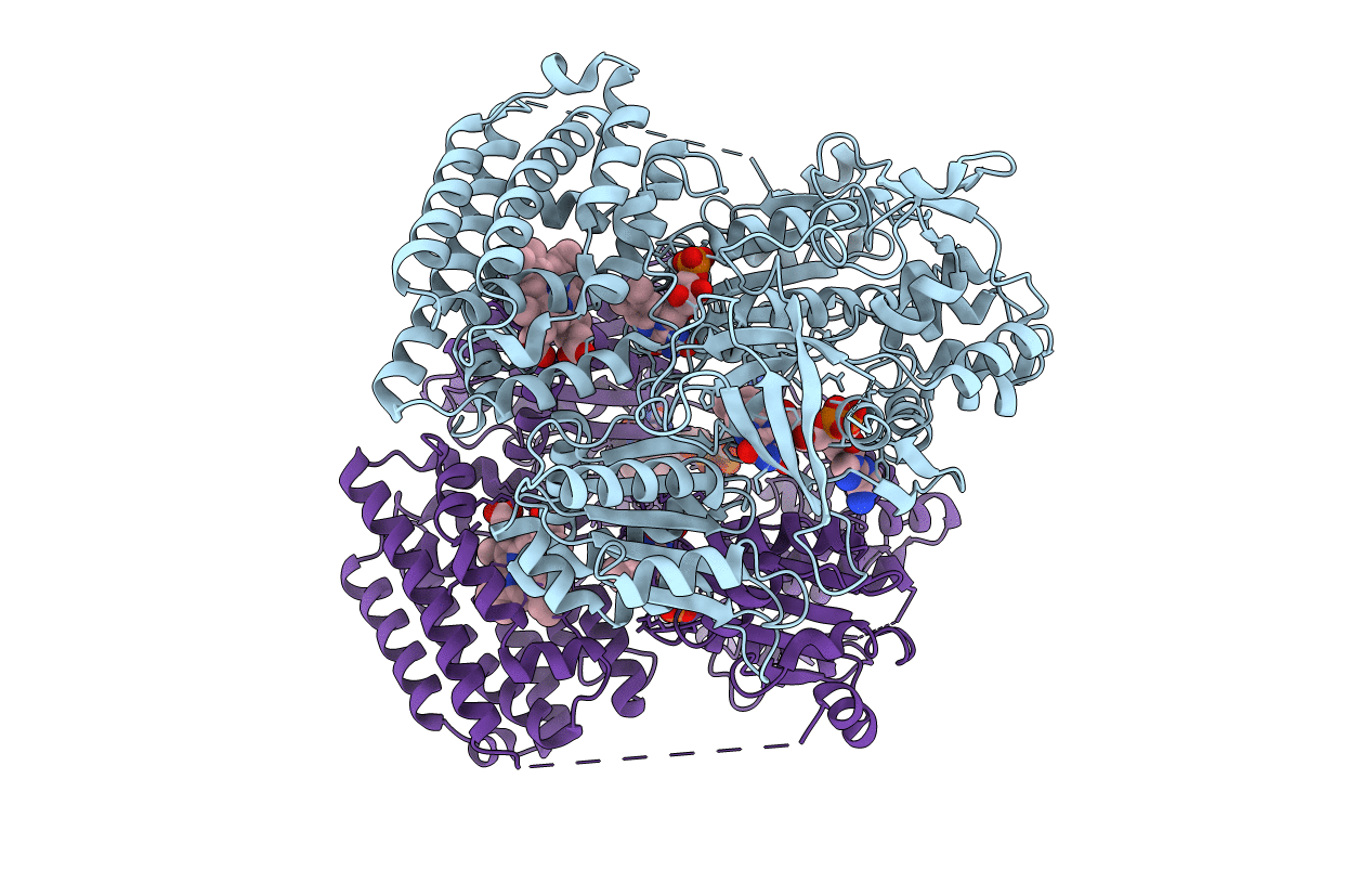
Fusion protein of heme oxygenase-1 and NADPH cytochrome P450 reductase (17aa)
Biological Source:
Source Organism:
Method Details:
Experimental Method:
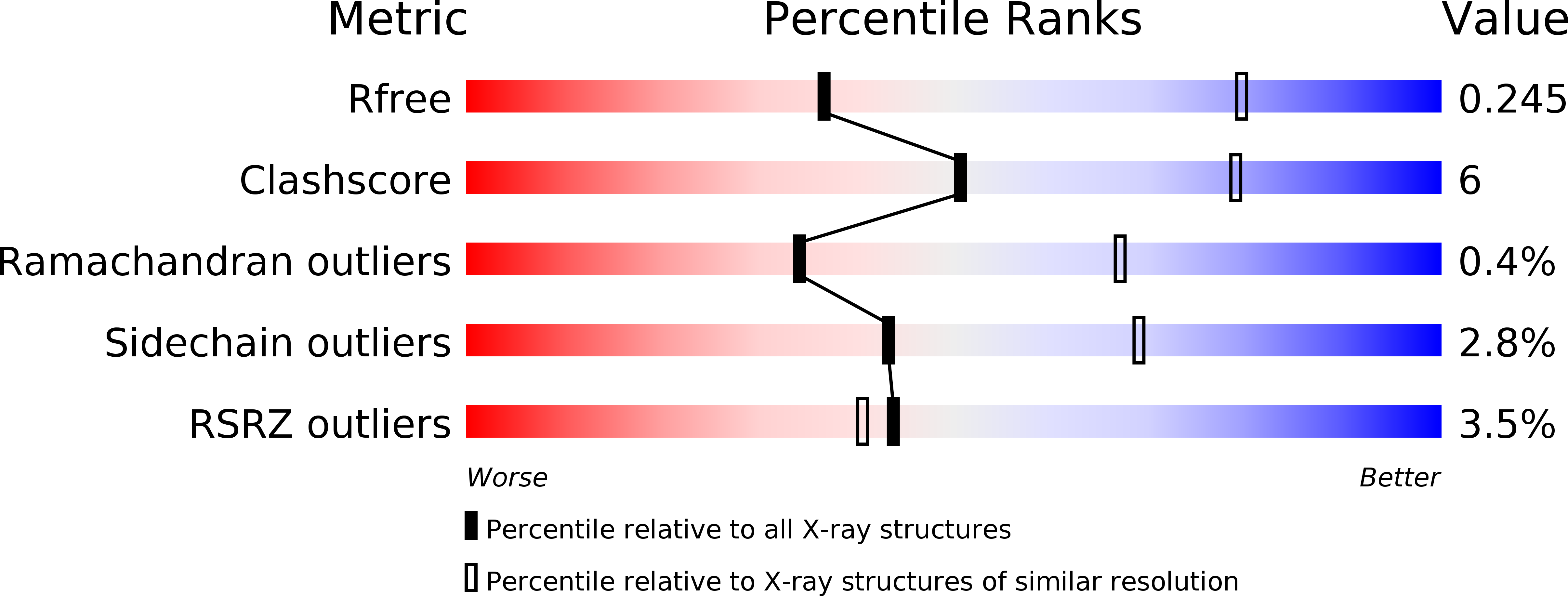
Resolution:
3.27 Å
R-Value Free:
0.24
R-Value Work:
0.22
R-Value Observed:
0.22
Space Group:
P 21 21 21
Macromolecular Entities
Polymer Type:polypeptide(L)
Molecule:Heme oxygenase 1,NADPH--cytochrome P450 reductase
Gene (Uniprot):Por, Hmox1
Mutations:T222P, P230A
Chain IDs:A, B
Chain Length:875
Number of Molecules:2
Biological Source:Rattus norvegicus
Primary Citation
Crystal structure of a NADPH-cytochrome P450 oxidoreductase (CYPOR) and heme oxygenase 1 fusion protein implies a conformational change in CYPOR upon NADPH/NADP+binding.
Febs Lett. 593 868 875 (2019)
PMID: 30883732 DOI: 10.1002/1873-3468.13360

Abstact

Heme oxygenase-1 (HMOX1) catalyzes heme degradation utilizing reducing equivalents supplied from NADPH-cytochrome P450 reductase (CYPOR). Recently, we determined the complex structure of NADP+ -bound open-conformation stabilized CYPOR and heme-HMOX1, but the resolution was limited to 4.3 Å. Here, we determined the crystal structure of the fusion protein of open-conformation stabilized CYPOR and heme-HMOX1 at 3.25 Å resolution. Unexpectedly, no NADP+ was bound to this fusion protein in the crystal. Structural comparison of the NADP+ -bound complex and the NADP+ -free fusion protein suggests that NADP+ binding regulates the conformational change in the FAD-binding domain of CYPOR. As a result of this change, the FMN-binding domain of CYPOR approaches heme-bound HMOX1 upon NADP+ binding to enhance the electron-transfer efficiency from FMN to heme.

Legend

Protein

Chemical

Disease

Primary Citation of related structures