6J66 image
Deposition Date 2019-01-14
Release Date 2019-07-10
Last Version Date 2024-11-06
Entry Detail
PDB ID:
6J66
Keywords:
Title:
Chondroitin sulfate/dermatan sulfate endolytic 4-O-sulfatase
Biological Source:
Source Organism:
Method Details:
Experimental Method:
Resolution:
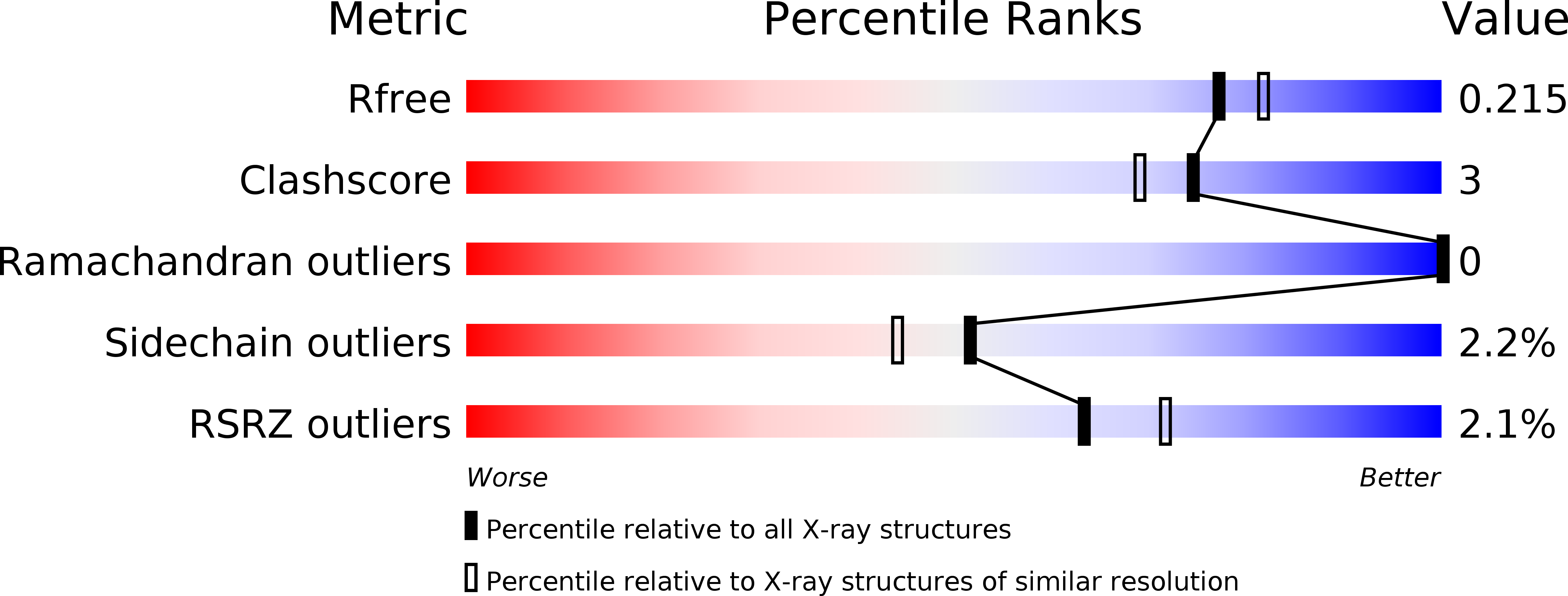
1.95 Å
R-Value Free:
0.21
R-Value Work:
0.18
R-Value Observed:
0.18
Space Group:
C 1 2 1
Macromolecular Entities
Structures with similar UniProt ID
Protein Blast
Polymer Type:polypeptide(L)
Molecule:Chondroitin sulfate/dermatan sulfate 4-O-endosulfatase protein
Mutagens:C96FGly
Chain IDs:A, B
Chain Length:521
Number of Molecules:2
Biological Source:Vibrio sp. FC509
Ligand Molecules
Primary Citation
Comparative Study of Two Chondroitin Sulfate/Dermatan Sulfate 4-O-Sulfatases With High Identity.
Front Microbiol 10 1309 1309 (2019)
PMID: 31244815 DOI: 10.3389/fmicb.2019.01309

Abstact

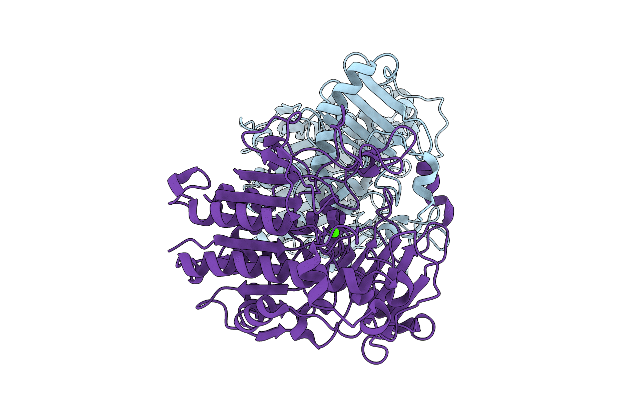
Chondroitin sulfate/dermatan sulfate (CS/DS) sulfatases are potential tools for structural and functional studies of CD/DS chains. In our previous study, a CS/DS 4-O-endosulfatase (endoVB4SF) was identified from a marine bacterium (Wang et al., 2015). Herein, another CS/DS 4-O-sulfatase (exoPB4SF) was identified from a Photobacterium sp. ExoPB4SF shares an 83% identity with endoVB4SF but showed strict exolytic activity. Comparative studies were performed for both enzymes on the basis of biochemical features, substrate-degrading patterns and three-dimensional structures. exoPB4SF exhibited a wider temperature and pH adaptability and better thermostability than endoVB4SF. Furthermore, exoPB4SF is a strict exolytic sulfatase that only releases the sulfate group from the GalNAc residue located at the reducing end, whereas endoVB4SF preferentially removed sulfate esters from the reducing end toward the non-reducing end though its directional degradation property was not strict. In addition, the structure of endoVB4SF was determined by X-ray crystallography at 1.95 Å. It adopts a globular conformation with two monomers per asymmetric unit. The exoPB4SF structure was constructed by homology modeling. Molecular docking results showed that although the residues around the catalytic center are conserved, the residues at the active site of endoVB4SF adopted a more favorable conformation for the binding of long CS/DS chains than those of exoPB4SF, which may explain why the two highly homogenous sulfatases possessed different action patterns. The results of this study provide insight into the structure-function relationship of CS/DS endo- and exosulfatases for the first time.

Legend

Protein

Chemical

Disease

Primary Citation of related structures