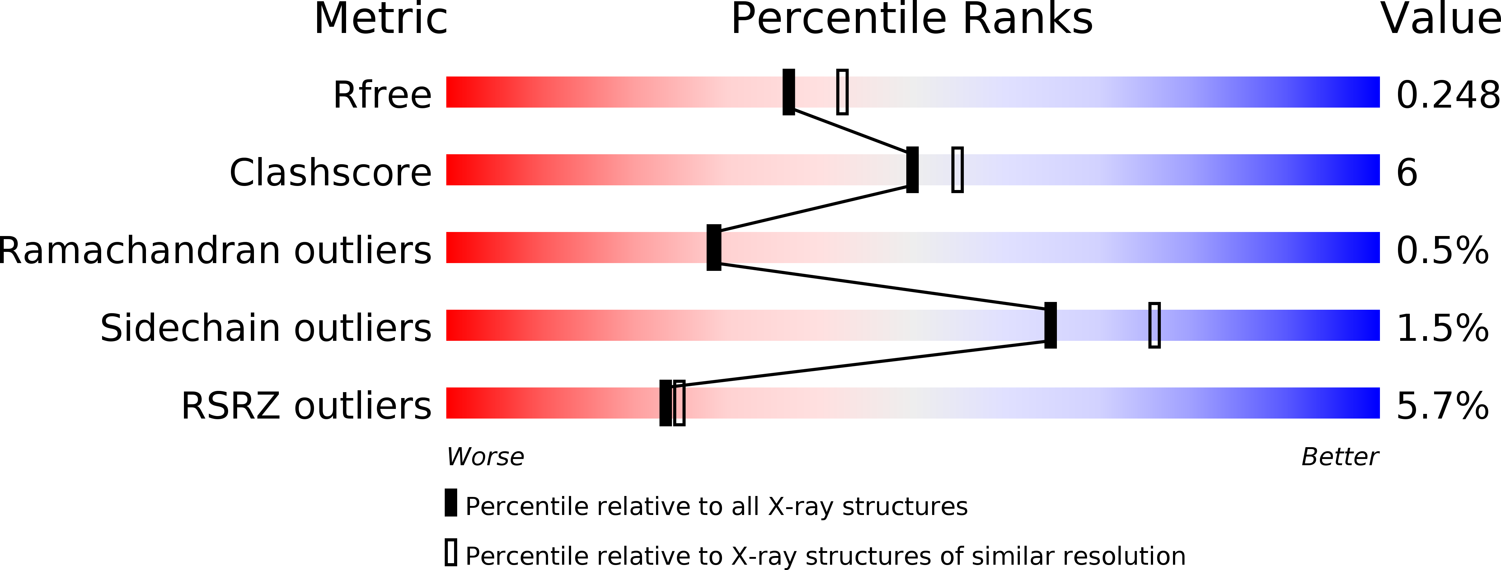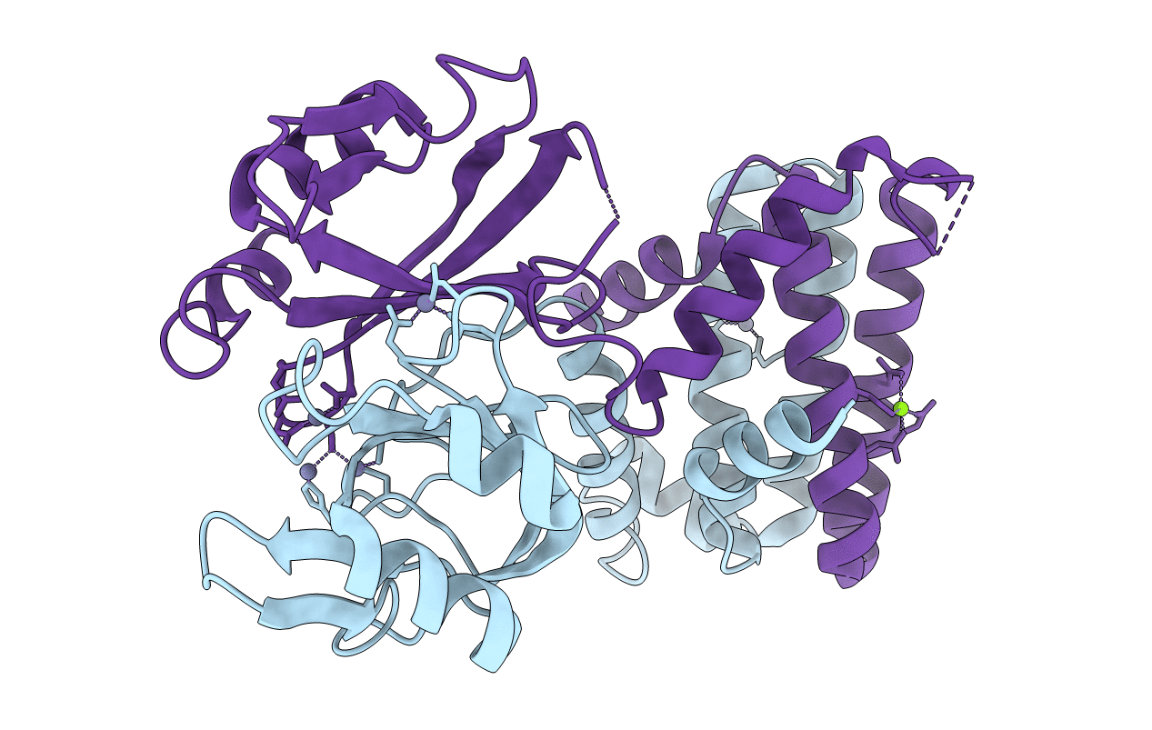
6J2L image
Deposition Date 2019-01-01
Release Date 2020-01-01
Last Version Date 2024-03-27
Entry Detail
PDB ID:
6J2L
Keywords:
Title:
Crystal structure of Bi-functional enzyme
Biological Source:
Source Organism:
Method Details:
Experimental Method:
Resolution:
2.17 Å
R-Value Free:
0.24
R-Value Work:
0.20
R-Value Observed:
0.20
Space Group:
C 1 2 1
Macromolecular Entities
Polymer Type:polypeptide(L)
Molecule:Histidine biosynthesis bifunctional protein HisIE
Gene (Uniprot):hisI
Chain IDs:A, B
Chain Length:202
Number of Molecules:2
Biological Source:Shigella flexneri
Primary Citation
Structural analysis of Shigella flexneri bi-functional enzyme HisIE in histidine biosynthesis.
Biochem.Biophys.Res.Commun. 516 540 545 (2019)
PMID: 31235255 DOI: 10.1016/j.bbrc.2019.06.099

Abstact

Histidine biosynthesis, which is absent in animals, was shown to be highly conserved among gram-negative bacteria, thus making it an attractive target for antibiotic design. There are many fusion forms of enzymes in the histidine biosynthetic pathway and people still have limited knowledge about their domain organizations and catalytic mechanisms, due to the lack of structural information. Here we report the first crystal structure of Shigella flexneri bi-functional enzyme HisIE (SfHisIE) that functions in the 2nd and 3rd steps in the histidine biosynthetic pathway. This structure shows that HisIE exists as dimers with two loops (fusion loop) connecting the individual dimer of HisE and HisI in its N-terminus and C-terminus respectively. Our mutagenesis study shows mutations in this fusion loop are lethal for bacteria indicating the advantage of gene fusion in Histidine biosynthesis. Structural analysis revealed several highly conserved residues in the putative ligand binding grooves of HisE and HisI, showing an evolutionarily conserved catalytic mechanism shared among gram negative-bacteria.

Legend

Protein

Chemical

Disease

Primary Citation of related structures