
Deposition Date
2018-12-28
Release Date
2019-11-06
Last Version Date
2024-03-27
Entry Detail
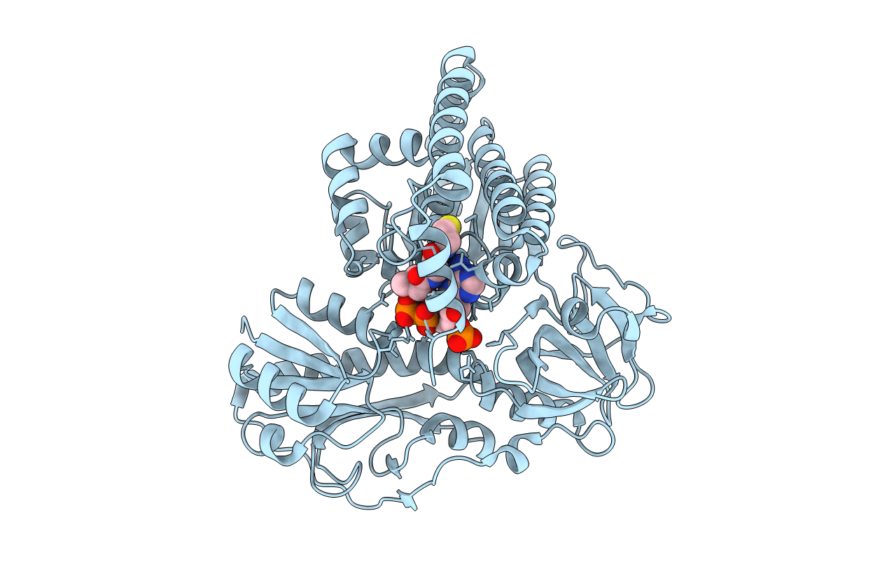
PDB ID:
6J1E
Keywords:
Title:
Crystal structure of HypX from Aquifex aeolicus (Crystal Form II)
Biological Source:
Source Organism:
Aquifex aeolicus VF5 (Taxon ID: 224324)
Host Organism:
Method Details:
Experimental Method:
Resolution:
2.40 Å
R-Value Free:
0.24
R-Value Work:
0.18
Space Group:
P 41 21 2
