6IZH image
Deposition Date 2018-12-19
Release Date 2019-01-02
Last Version Date 2023-11-22
Entry Detail
PDB ID:
6IZH
Keywords:
Title:
Crystal structure of deaminase AmnE from Pseudomonas sp. AP-3
Biological Source:
Source Organism:
Pseudomonas sp (Taxon ID: 306)
Method Details:
Experimental Method:
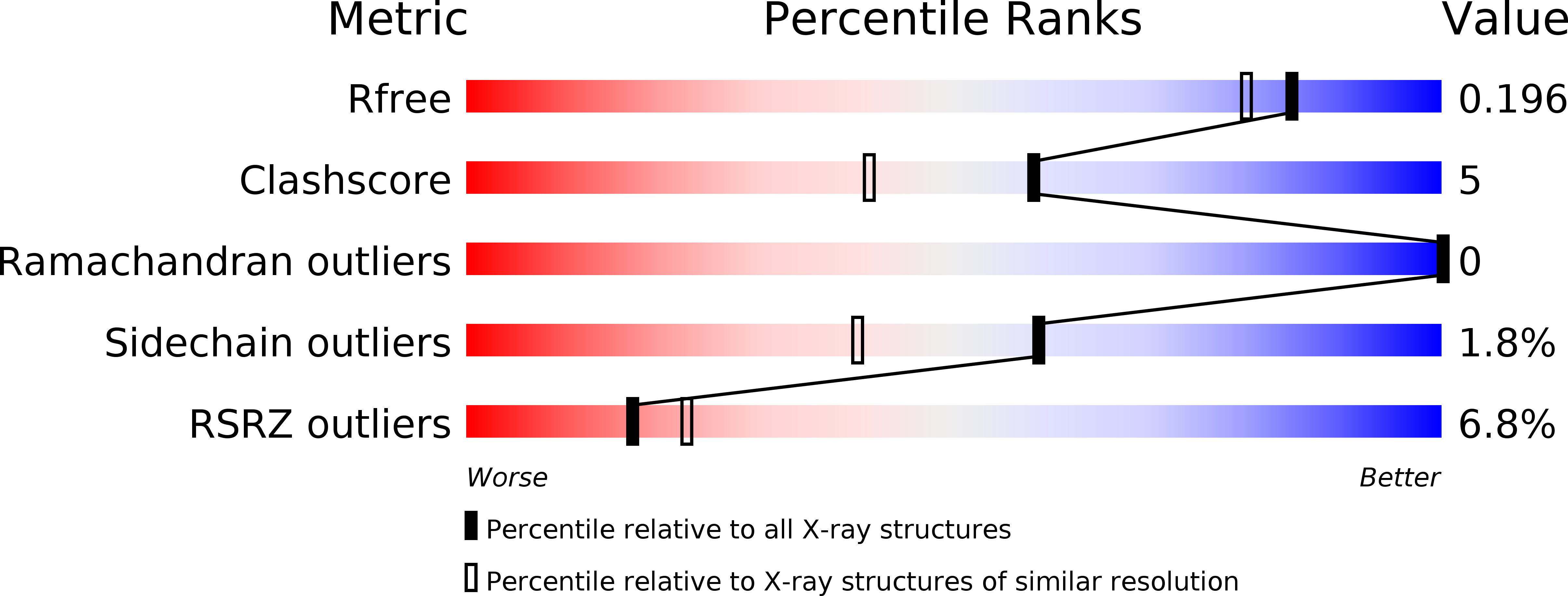
Resolution:
1.75 Å
R-Value Free:
0.19
R-Value Work:
0.15
R-Value Observed:
0.15
Space Group:
C 1 2 1
Macromolecular Entities
Structures with similar UniProt ID
Protein Blast
Polymer Type:polypeptide(L)
Molecule:2-aminomuconate deaminase
Gene (Uniprot):amnD
Chain IDs:A, B, C, D, E, F, G, H, I
Chain Length:142
Number of Molecules:9
Biological Source:Pseudomonas sp
Ligand Molecules
Primary Citation
A Unique Homo-Hexameric Structure of 2-Aminomuconate Deaminase in the BacteriumPseudomonas species AP-3.
Front Microbiol 10 2079 2079 (2019)
PMID: 31555255 DOI: 10.3389/fmicb.2019.02079

Abstact

The bacterium Pseudomonas species sp. AP-3 is one of several microorganisms that are capable of using 2-aminophenol as its sole source of carbon, nitrogen and energy. Several 2-aminophenol-metabolizing enzymes have pivotal roles in the biodegradation of aniline and its derivatives as environmental pollutants in Pseudomonas. The bacterium Pseudomonas sp. AP-3 recruits a unique 2-aminomuconate deaminase (AmnE) to hydrolyze 2-aminomuconate to 4-oxalocrotonate, and releases ammonia in the modified meta-cleavage pathway by forming various compounds-including acetaldehyde, pyruvic acid, acetyl-CoA, and succinate-that may enter the Krebs cycle. AmnE also belongs to the YjgF/YER057c/UK114 family (also known as the Rid family), which is conserved in all domains of life and prefers structurally homotrimeric forms with diverse functional purposes. To study the mechanism of the modified meta-cleavage pathway in Pseudomonas sp. AP-3, we determined the first crystal structure of AmnE from Pseudomonas sp. AP-3 at 1.75 Å. AmnE forms a unique homohexamer instead of a trimer which is normally adopted by the members of YjgF/YER057c/UK114 family. Based on the structure of the AmnE hexamer, we observed a hydrophobic base composed of six Lp3 loops (residues 122-131) in each of the AmnE protomers that have pivotal roles in the assembly of the hexamer. Eighteen hydrogen bonds formed by the residues Met96, Pro126, and Arg56, which surround the hydrophobic base, allowed the combination of the two trimers into a stable hexamer. The single mutant of AmnE R56A lost the ability to maintain the hexameric conformation, and revealed that the hydrogen bonds between residues Arg56 and Met96 have pivotal roles in the AmnE hexameric assembly.

Legend

Protein

Chemical

Disease

Primary Citation of related structures