
Deposition Date
2018-12-18
Release Date
2019-05-01
Last Version Date
2023-11-22
Entry Detail
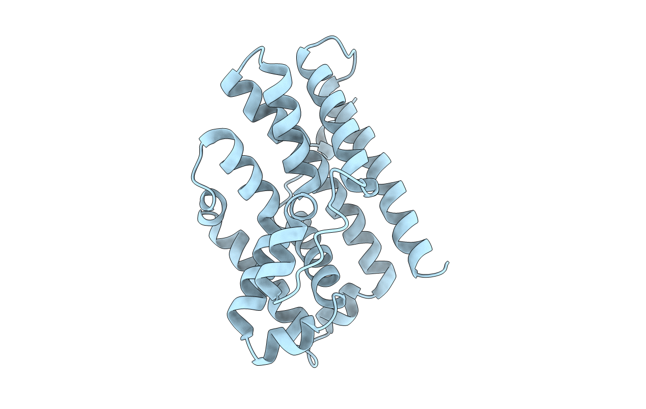
PDB ID:
6IYZ
Keywords:
Title:
Structural basis for activity of TRIC counter-ion channels in calcium release
Biological Source:
Source Organism:
Gallus gallus (Taxon ID: 9031)
Host Organism:
Method Details:
Experimental Method:
Resolution:
2.20 Å
R-Value Free:
0.27
R-Value Work:
0.25
R-Value Observed:
0.25
Space Group:
F 41 3 2
