6IWY image
Deposition Date 2018-12-08
Release Date 2019-05-22
Last Version Date 2024-03-27
Entry Detail
PDB ID:
6IWY
Title:
Crystal structure of the flagellar cap protein FliD from Helicobacter pylori
Biological Source:
Method Details:
Experimental Method:
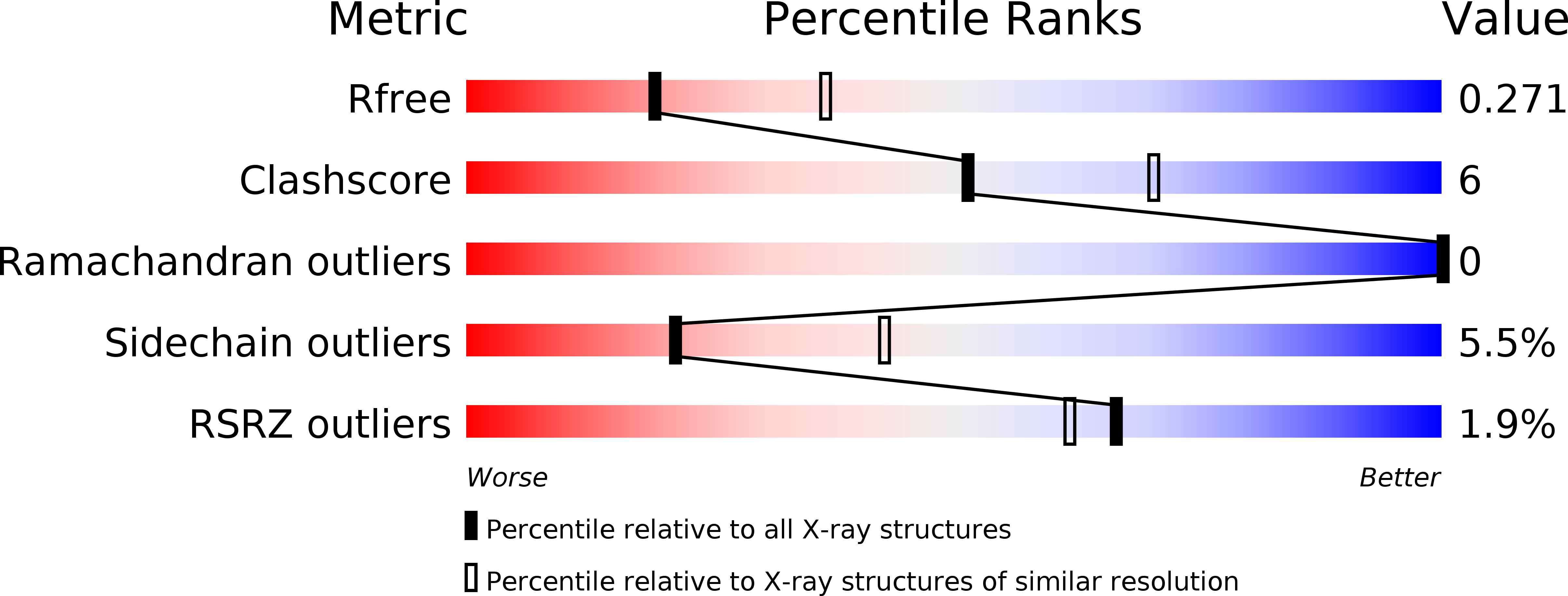
Resolution:
2.60 Å
R-Value Free:
0.27
R-Value Work:
0.23
R-Value Observed:
0.24
Space Group:
P 2 2 21
Macromolecular Entities
Structures with similar UniProt ID
Protein Blast
Polymer Type:polypeptide(L)
Molecule:Flagellar hook-associated protein 2
Gene (Uniprot):fliD
Chain IDs:A
Chain Length:412
Number of Molecules:1
Biological Source:Helicobacter pylori (strain ATCC 700392 / 26695)
Primary Citation
Structural analysis of the flagellar capping protein FliD from Helicobacter pylori.
Biochem.Biophys.Res.Commun. 514 98 104 (2019)
PMID: 31023530 DOI: 10.1016/j.bbrc.2019.04.065

Abstact

Helicobacter pylori is a pathogenic flagellated bacterium that infects the gastroduodenal mucosa and causes peptic ulcers in humans. FliD caps the distal end of the flagellar filament and is essential in filament growth. Moreover, FliD has been studied to diagnose and prevent H. pylori infection. Here, we report structure-based molecular studies of H. pylori FliD (hpFliD). A crystal structure of hpFliD at 2.6 Å resolution presents a four-domain (D2-D5) structure, where the D3 domain forms a central platform surrounded by the other three domains (D2, D4, and D5). hpFliD domains D2 and D3 structurally resemble those of FliD orthologs, whereas the D4 and D5 domains are exclusive to hpFliD. Moreover, our ELISA analysis using anti-H. pylori antibodies demonstrated that the hpFliD-specific D4 and D5 domains are highly antigenic compared to the D2 and D3 domains. Collectively, our structural and serological analyses underscore the structural role of hpFliD domains and provide a molecular basis for vaccine and diagnosis development.

Legend

Protein

Chemical

Disease

Primary Citation of related structures