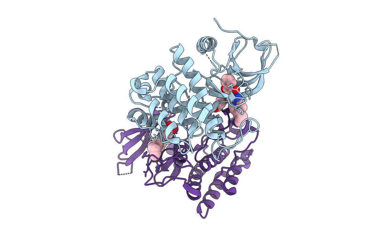
6IQN image
Deposition Date 2018-11-08
Release Date 2020-01-22
Last Version Date 2023-11-22
Entry Detail
PDB ID:
6IQN
Keywords:
Title:
Crystal structure of TrkA kinase with ligand
Biological Source:
Source Organism:
Homo sapiens (Taxon ID: 9606)
Host Organism:
Method Details:
Experimental Method:
Resolution:
2.54 Å
R-Value Free:
0.28
R-Value Work:
0.22
R-Value Observed:
0.22
Space Group:
P 41 21 2
Macromolecular Entities
Polymer Type:polypeptide(L)
Molecule:High affinity nerve growth factor receptor
Gene (Uniprot):NTRK1
Chain IDs:A, B
Chain Length:296
Number of Molecules:2
Biological Source:Homo sapiens
Ligand Molecules
Primary Citation
An isoform-selective inhibitor of tropomyosin receptor kinase A behaves as molecular glue.
Bioorg.Med.Chem.Lett. 30 126775 126775 (2020)
PMID: 31699609 DOI: 10.1016/j.bmcl.2019.126775

Abstact

The production of TrkA-selective inhibitors is considerably difficult because the kinase domains of TrkA and its isoforms TrkB/C have highly homologous amino acid sequences. Here we describe the structural basis for the acquisition of selectivity for a isoform-selective TrkA inhibitor, namely compound V1. The X-ray structure revealed that V1 acts as a molecular glue to stabilize the symmetrical dimer of the TrkA kinase domains. V1 binds to the ATP-binding site and simultaneously engages in the dimeric interface of TrkA. The region of the dimeric interface in TrkA is not conserved in TrkB/C; thus, dimer formation may be a novel mechanism for the production of selective TrkA inhibitors. The biochemical and biophysical assay results confirmed that V1 selectively inhibited TrkA and induced the dimer formation of TrkA, but not TrkB. The binding pocket at the TrkA dimer interface can be used for the production of new isoform-selective TrkA inhibitors.

Legend

Protein

Chemical

Disease

Primary Citation of related structures