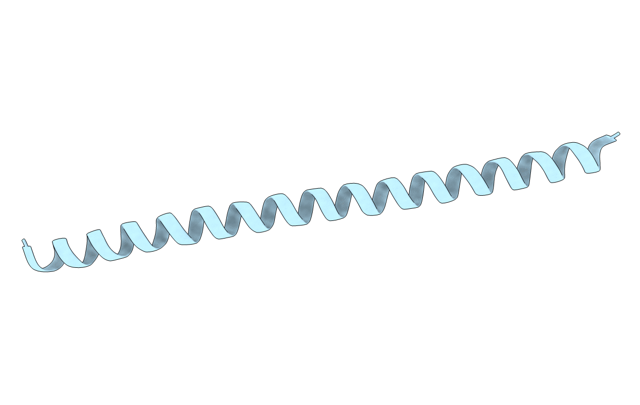
6IQE image
Entry Detail
PDB ID:
6IQE
Keywords:
Title:
Human prohibitin 2
Biological Source:
Source Organism:
Host Organism:
PDB Version:
Deposition Date:
2018-11-07
Release Date:
2019-09-25
Method Details:
Experimental Method:
Resolution:
1.70 Å
R-Value Free:
0.23
R-Value Work:
0.21
R-Value Observed:
0.21
Space Group:
P 41 2 2
Macromolecular Entities
Polymer Type:polypeptide(L)
Description:Prohibitin-2
Chain IDs:A
Chain Length:87
Number of Molecules:1
Biological Source:Homo sapiens
Primary Citation
Structural Basis of Mitochondrial Scaffolds by Prohibitin Complexes: Insight into a Role of the Coiled-Coil Region.
Iscience 19 1065 1078 (2019)
PMID: 31522117 DOI: 10.1016/j.isci.2019.08.056

Abstact

The coiled-coil motif mediates subunit oligomerization and scaffolding and underlies several fundamental biologic processes. Prohibitins (PHBs), mitochondrial inner membrane proteins involved in mitochondrial homeostasis and signal transduction, are predicted to have a coiled-coil motif, but their structural features are poorly understood. Here we solved the crystal structure of the heptad repeat (HR) region of PHB2 at 1.7-Å resolution, showing that it assembles into a dimeric, antiparallel coiled-coil with a unique negatively charged area essential for the PHB interactome in mitochondria. Disruption of the HR coiled-coil abolishes well-ordered PHB complexes and the mitochondrial tubular networks accompanying PHB-dependent signaling. Using a proximity-dependent biotin identification (BioID) technique in live cells, we mapped a number of mitochondrial intermembrane space proteins whose association with PHB2 relies on the HR coiled-coil region. Elucidation of the PHB complex structure in mitochondria provides insight into essential PHB interactomes required for mitochondrial dynamics as well as signal transduction.

Legend

Protein

Chemical

Disease

Primary Citation of related structures