Deposition Date
2018-11-01
Release Date
2019-04-24
Last Version Date
2024-03-27
Entry Detail
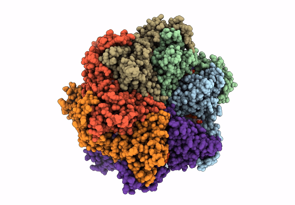
Biological Source:
Source Organism(s):
Cricetulus griseus (Taxon ID: 10029)
Expression System(s):
Method Details:
Experimental Method:
Resolution:
3.70 Å
Aggregation State:
PARTICLE
Reconstruction Method:
SINGLE PARTICLE