Deposition Date
1992-11-25
Release Date
1994-01-31
Last Version Date
2024-10-16
Entry Detail
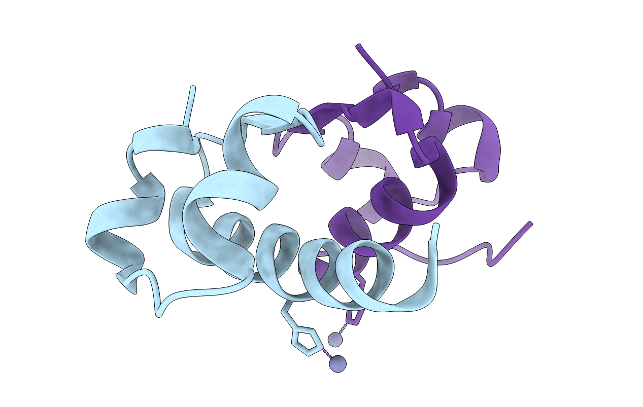
PDB ID:
6INS
Keywords:
Title:
X-RAY ANALYSIS OF THE SINGLE CHAIN B29-A1 PEPTIDE-LINKED INSULIN MOLECULE. A COMPLETELY INACTIVE ANALOGUE
Biological Source:
Source Organism(s):
Sus scrofa (Taxon ID: 9823)
Method Details: