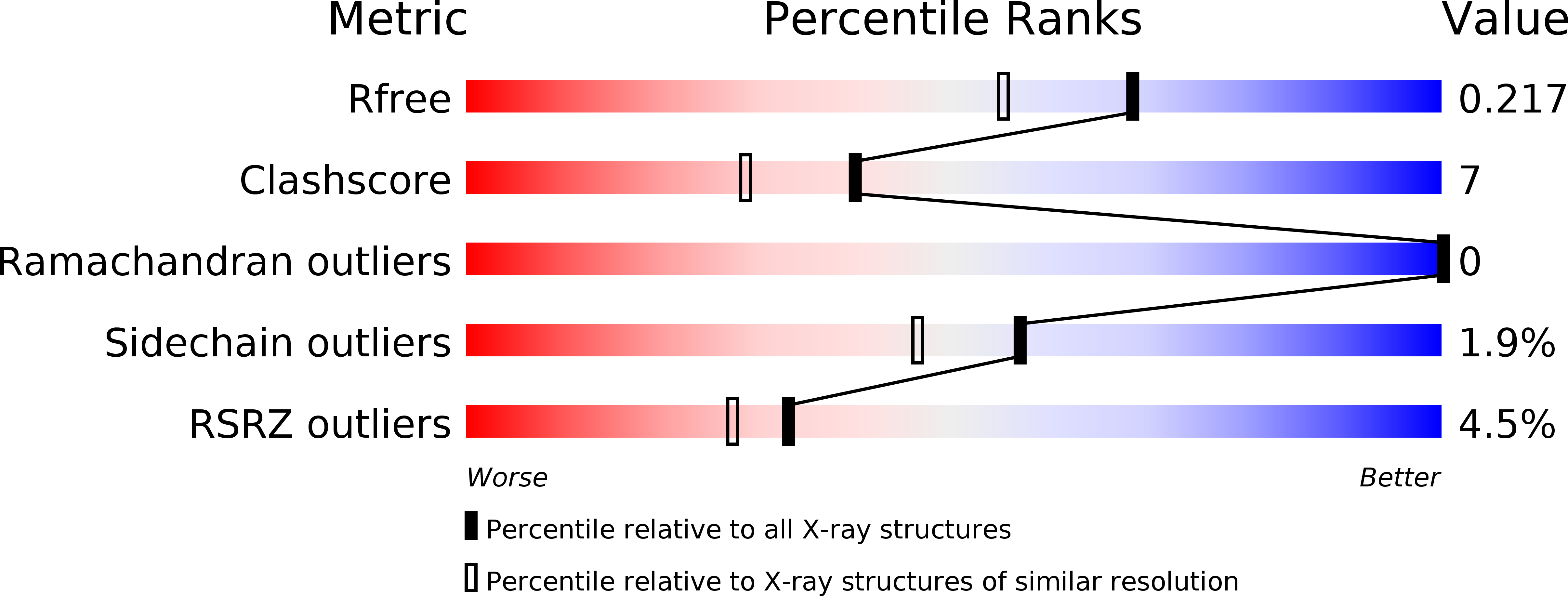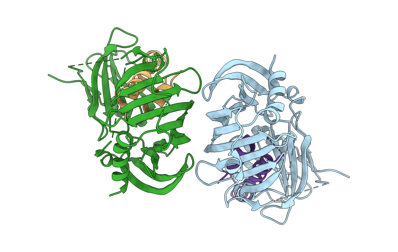
6IN9 image
Entry Detail
PDB ID:
6IN9
Title:
Crystal structure of MucB in complex with MucA(peri)
Biological Source:
PDB Version:
Deposition Date:
2018-10-24
Release Date:
2019-07-24
Method Details:
Experimental Method:
Resolution:
1.80 Å
R-Value Free:
0.21
R-Value Work:
0.18
R-Value Observed:
0.18
Space Group:
P 1 21 1
Macromolecular Entities
Polymer Type:polypeptide(L)
Description:Sigma factor AlgU regulatory protein MucB
Chain IDs:A, B
Chain Length:295
Number of Molecules:2
Biological Source:Pseudomonas aeruginosa (strain ATCC 15692 / DSM 22644 / CIP 104116 / JCM 14847 / LMG 12228 / 1C / PRS 101 / PAO1)
Polymer Type:polypeptide(L)
Description:Sigma factor AlgU negative regulatory protein
Chain IDs:C, D
Chain Length:89
Number of Molecules:2
Biological Source:Pseudomonas aeruginosa (strain ATCC 15692 / DSM 22644 / CIP 104116 / JCM 14847 / LMG 12228 / 1C / PRS 101 / PAO1)
Primary Citation
Structural basis for the recognition of MucA by MucB and AlgU in Pseudomonas aeruginosa.
Febs J. 286 4982 4994 (2019)
PMID: 31297938 DOI: 10.1111/febs.14995

Abstact

Alginate production in Pseudomonas aeruginosa is regulated by the alternate σ factor AlgU, which in turn is regulated by the MucABCD system. The anti-σ factor MucA binds AlgU in the cytoplasm and prevents AlgU from binding to the RNA polymerase for transcription. MucB binds MucA in the periplasm and inhibits proteolysis of MucA and subsequent release of AlgU. In this work, we report crystal structures of MucA in complex with AlgU and MucB. A structure of MucB alone reveals the structural changes required for MucA recognition. A unique disulfide bond is identified in MucB, and mutation of this disulfide bond results in a shift from monomer to MucB dimers or tetramers. As MucB tetramers have previously been shown to be unable to bind MucA, this suggests a redox-sensitive stress response mechanism in MucB. The AlgU-MucA structure reveals a conserved σ factor/anti-σ factor complex, but AlgU lacks a disulfide bond conserved in many other σ factors. Our structures reveal the molecular basis for MucA recognition by MucB in the periplasm and AlgU in the cytoplasm, thus providing an important step in understanding the mechanisms that regulate a key signal transduction pathway involved in P. aeruginosa pathogenesis. DATABASE: The atomic coordinates and structure factors for MucAcyto -AlgU, MucB, and MucAperi -MucB have been deposited in the Protein Data Bank (PDB) with the accession code 6IN7, 6IN8, and 6IN9, respectively.

Legend

Protein

Chemical

Disease

Primary Citation of related structures