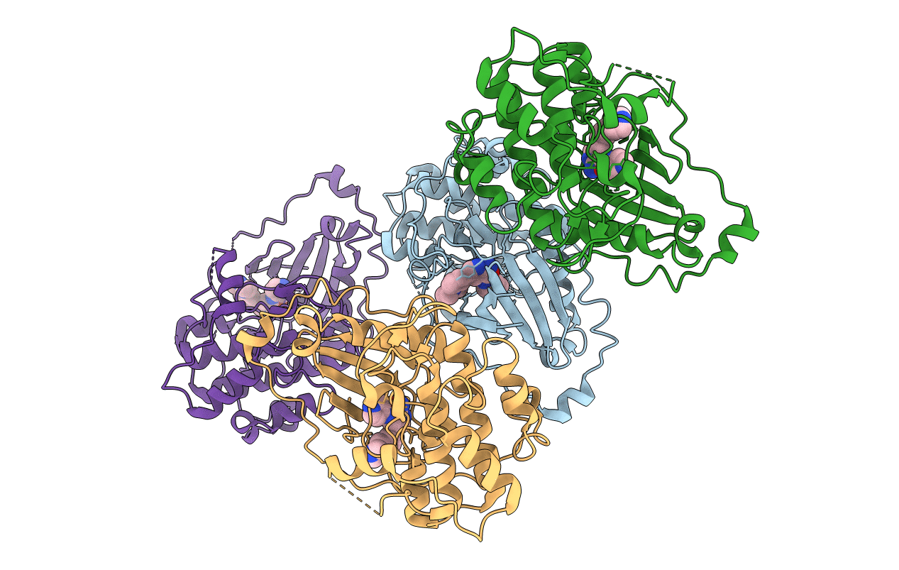
6ILZ image
Entry Detail
PDB ID:
6ILZ
Keywords:
Title:
Crystal structure of PKCiota in complex with inhibitor
Biological Source:
Source Organism:
Host Organism:
PDB Version:
Deposition Date:
2018-10-21
Release Date:
2019-06-26
Method Details:
Experimental Method:
Resolution:
3.26 Å
R-Value Free:
0.34
R-Value Work:
0.30
R-Value Observed:
0.30
Space Group:
P 1 21 1
Macromolecular Entities
Polymer Type:polypeptide(L)
Description:Protein kinase C iota type
Chain IDs:A, B (auth: C), C (auth: E), D (auth: G)
Chain Length:345
Number of Molecules:4
Biological Source:Homo sapiens
Modified Residue
Compound ID Chain ID Parent Comp ID Details 2D Image
TPO A THR modified residue
Ligand Molecules
Primary Citation
Fragment-based Discovery of a Small-Molecule Protein Kinase C-iota Inhibitor Binding Post-kinase Domain Residues.
Acs Med.Chem.Lett. 10 318 323 (2019)
PMID: 30891133 DOI: 10.1021/acsmedchemlett.8b00546

Abstact

The atypical protein kinase C-iota (PKC-ι) enzyme is implicated in various cancers and has been put forward as an attractive target for developing anticancer therapy. A high concentration biochemical screen identified pyridine fragment weakly inhibiting PKC-ι with IC50 = 424 μM. Driven by structure-activity relationships and guided by docking hypothesis, the weakly bound fragment was eventually optimized into a potent inhibitor of PKC-ι (IC50= 270 nM). Through the course of the optimization, an intermediate compound was crystallized with the protein, and careful analysis of the X-ray crystal structure revealed a unique binding mode involving the post-kinase domain (C-terminal tail) of PKC-ι.

Legend

Protein

Chemical

Disease

Primary Citation of related structures