
Deposition Date
2018-10-17
Release Date
2019-07-24
Last Version Date
2024-10-23
Entry Detail
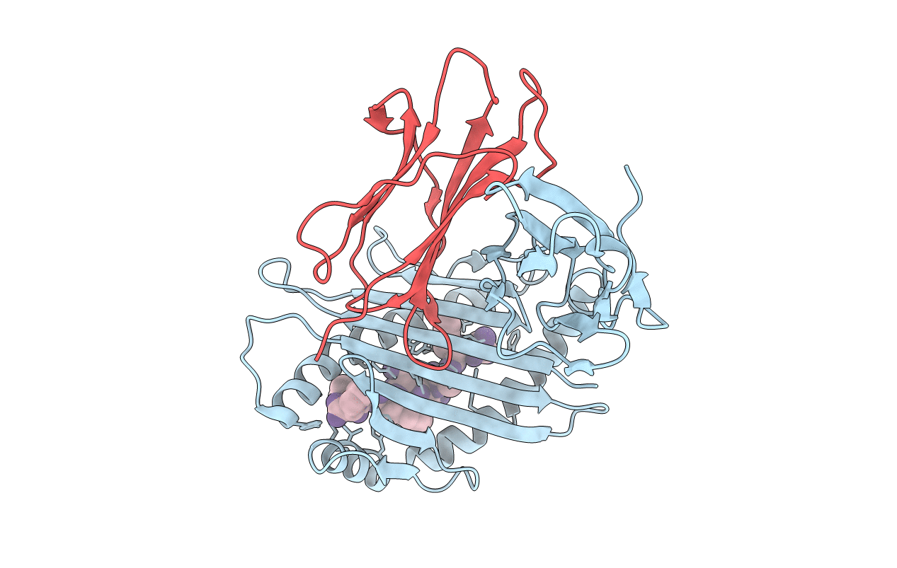
PDB ID:
6ILE
Keywords:
Title:
CRYSTAL STRUCTURE OF A MUTANT PTAL-N*01:01 FOR 2.9 ANGSTROM, 52M 53D 54L DELETED
Biological Source:
Source Organism(s):
Pteropus alecto (Taxon ID: 9402)
Hendra virus (Taxon ID: 63330)
Hendra virus (Taxon ID: 63330)
Expression System(s):
Method Details:
Experimental Method:
Resolution:
2.90 Å
R-Value Free:
0.29
R-Value Work:
0.21
R-Value Observed:
0.21
Space Group:
C 2 2 21
