6IKI image
Entry Detail
PDB ID:
6IKI
Title:
Crystal structure of YfiB(W55L)
Biological Source:
PDB Version:
Deposition Date:
2018-10-16
Release Date:
2019-03-13
Method Details:
Experimental Method:
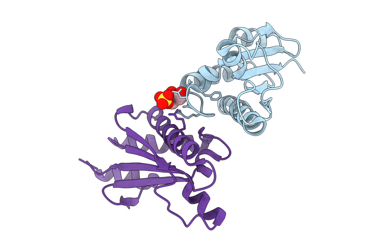
Resolution:
2.20 Å
R-Value Free:
0.24
R-Value Work:
0.20
R-Value Observed:
0.20
Space Group:
P 41
Macromolecular Entities
Polymer Type:polypeptide(L)
Description:YfiB
Mutations:W55L
Chain IDs:A, B
Chain Length:168
Number of Molecules:2
Biological Source:Pseudomonas aeruginosa (strain ATCC 15692 / DSM 22644 / CIP 104116 / JCM 14847 / LMG 12228 / 1C / PRS 101 / PAO1)
Primary Citation
Structural analysis of activating mutants of YfiB from Pseudomonas aeruginosa PAO1.
Biochem. Biophys. Res. Commun. 506 997 1003 (2018)
PMID: 30404734 DOI: 10.1016/j.bbrc.2018.10.190

Abstact

Bacterial cyclic-di-GMP (c-di-GMP) is an important messenger molecule that influences diverse cellular processes including motility, virulence and cytotoxicity systems, polysaccharide synthesis and biofilm formation. The YfiBNR tripartite signalling system in P. aeruginosa modulates the cellular c-di-GMP levels in response to signals received from the periplasm. In this study, we analyse the structures of activating mutants of the outer membrane protein YfiB that give rise to increased surface attachment and biofilm formation. The F48S and W55L mutants of YfiB(27-168) crystallize in the same dimeric arrangement as our previously reported YfiB structures that preclude complex formation with YfiR. The L43P mutant of YfiB(27-168) is monomeric and forms a stable complex with YfiR. The YfiB(L43P)-YfiR crystal structure reveals a dramatic rearrangement of the N-terminal fragment, which is implicated in increased YfiB activation and membrane attachment, upon YfiR binding. Comparison with our previous complex structure between YfiB(59-168) and YfiR reveals extensive interactions between the N-terminal fragment of YfiB (residues 35-55) and YfiR.

Legend

Protein

Chemical

Disease

Primary Citation of related structures