Deposition Date
2018-09-28
Release Date
2019-10-02
Last Version Date
2023-11-22
Entry Detail
PDB ID:
6IH7
Keywords:
Title:
Crystal structure of a standalone versatile EAL protein from Vibrio cholerae O395 - 3',3'-cGAMP bound form
Biological Source:
Source Organism(s):
Expression System(s):
Method Details:
Experimental Method:
Resolution:
2.25 Å
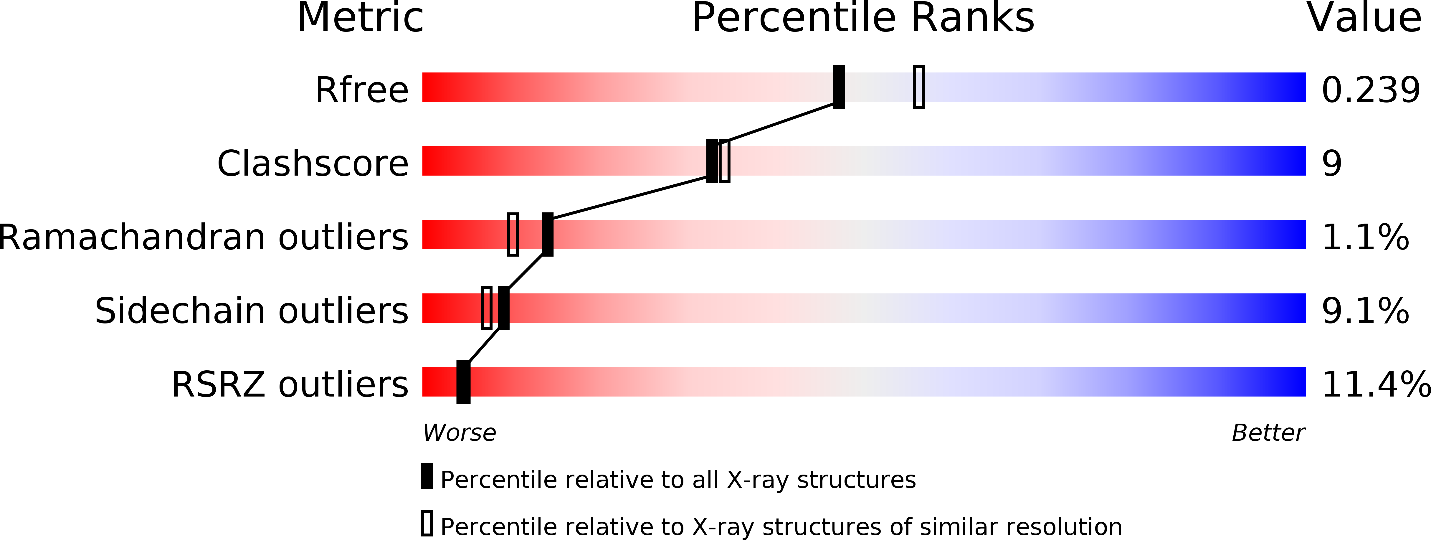
R-Value Free:
0.23
R-Value Work:
0.18
R-Value Observed:
0.18
Space Group:
C 1 2 1