6IGA image
Deposition Date 2018-09-25
Release Date 2019-02-06
Last Version Date 2023-11-22
Entry Detail
PDB ID:
6IGA
Keywords:
Title:
Crystal structure of argininosuccinate lyase from Mycobacterium tuberculosis
Biological Source:
Method Details:
Experimental Method:
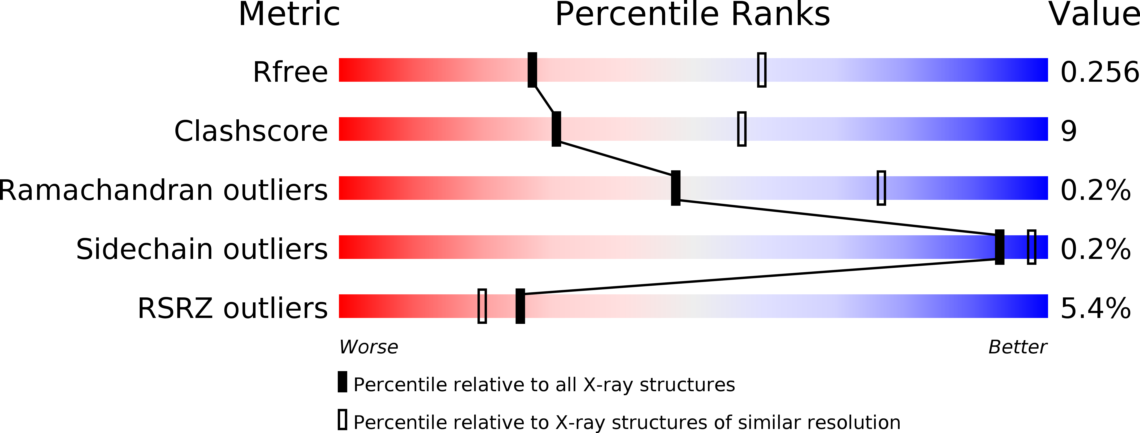
Resolution:
2.78 Å
R-Value Free:
0.25
R-Value Work:
0.21
R-Value Observed:
0.22
Space Group:
P 31 2 1
Macromolecular Entities
Structures with similar UniProt ID
Protein Blast
Polymer Type:polypeptide(L)
Molecule:Argininosuccinate lyase
Gene (Uniprot):argH
Chain IDs:A, B, C, D
Chain Length:470
Number of Molecules:4
Biological Source:Mycobacterium tuberculosis (strain ATCC 25618 / H37Rv)
Ligand Molecules
Primary Citation
Crystal structure and biochemical study on argininosuccinate lyase from Mycobacterium tuberculosis.
Biochem. Biophys. Res. Commun. 510 116 121 (2019)
PMID: 30665717 DOI: 10.1016/j.bbrc.2019.01.061

Abstact

Argininosuccinate lyase (ASL) participates in arginine synthesis through catalysing a reversible reaction in which argininosuccinate (AS) converts into arginine and fumarate. ASL from Mycobacterium tuberculosis is essential for its growth. In this work, the crystal structure of the apo form of MtbASL was determined and reveals a tetrameric structure that is essential for its activity since the active sites are formed by residues from three different monomers. Subsequently, we determined the crystal structure of MtbASL-sulfate complex, and the ligand mimics the negatively charged intermediate. The complex structure and mutagenesis studies indicate that residues S282 and H161 might act as a catalytic dyad. A major conformational change in the MtbASL-SO4 complex structure could be observed upon sulfate binding, and this movement facilitates the interaction between substrate and the residues involved in catalysis. A different conformational change in the C-terminal domain could be observed in the MtbASL-SO4 complex compared with that in other homologues. This difference may be responsible for the lower activity of MtbASL, which is related to the slow growth rate of M. tuberculosis. The C-terminal domain is a potential allosteric site upon inhibitor binding. The various conformational changes and the diversity of the sequence of the potential allosteric site across the homologues might provide clues for designing selective inhibitors against M. tuberculosis.

Legend

Protein

Chemical

Disease

Primary Citation of related structures
Feedback Form
Name
Email
Institute
Feedback