Deposition Date
2018-09-20
Release Date
2020-01-29
Last Version Date
2024-03-27
Entry Detail
PDB ID:
6IFM
Keywords:
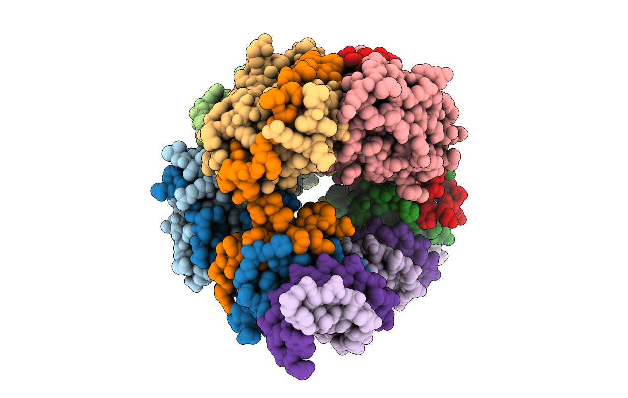
Title:
Crystal structure of DNA bound VapBC from Salmonella typhimurium
Biological Source:
Source Organism(s):
Salmonella enterica subsp. enterica serovar Typhimurium str. LT2 (Taxon ID: 99287)
synthetic construct (Taxon ID: 32630)
synthetic construct (Taxon ID: 32630)
Expression System(s):
Method Details:
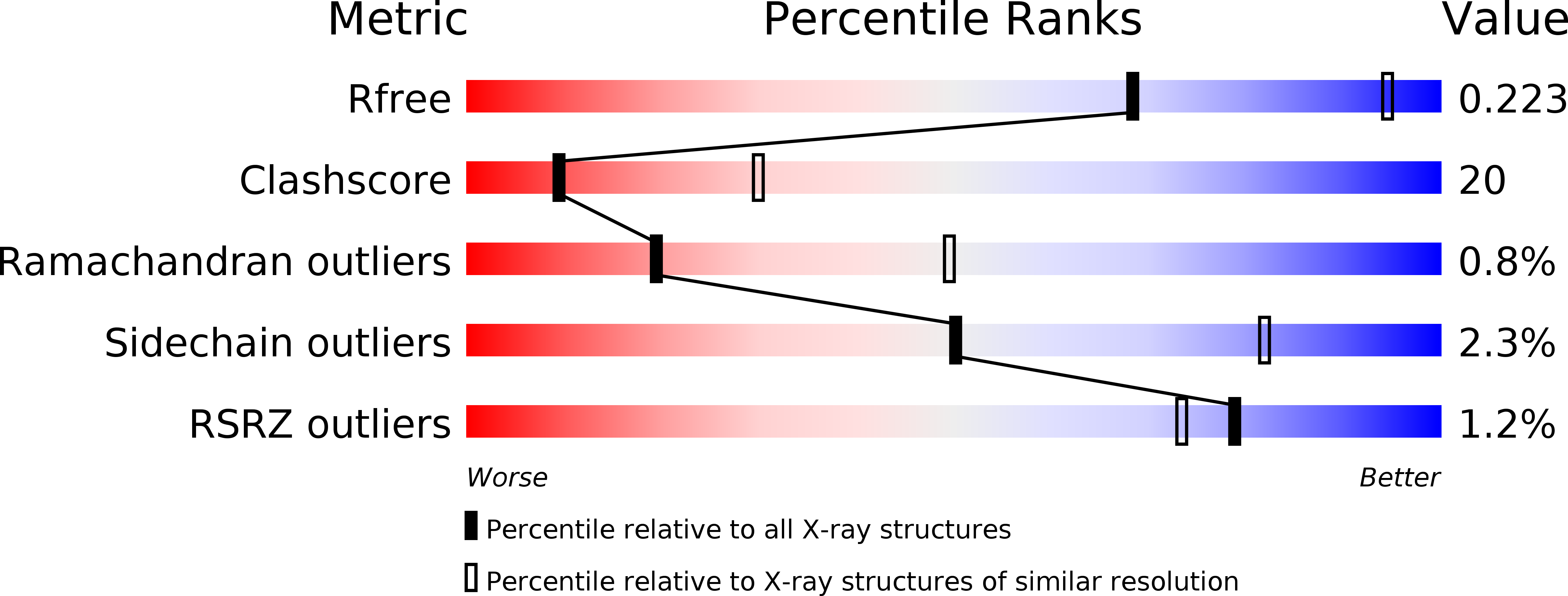
Experimental Method:
Resolution:
2.80 Å
R-Value Free:
0.23
R-Value Work:
0.20
R-Value Observed:
0.21
Space Group:
P 31