6IF6 image
Deposition Date 2018-09-18
Release Date 2019-05-01
Last Version Date 2024-03-27
Entry Detail
PDB ID:
6IF6
Title:
Structure of the periplasmic domain of SflA
Biological Source:
Source Organism:
Method Details:
Experimental Method:
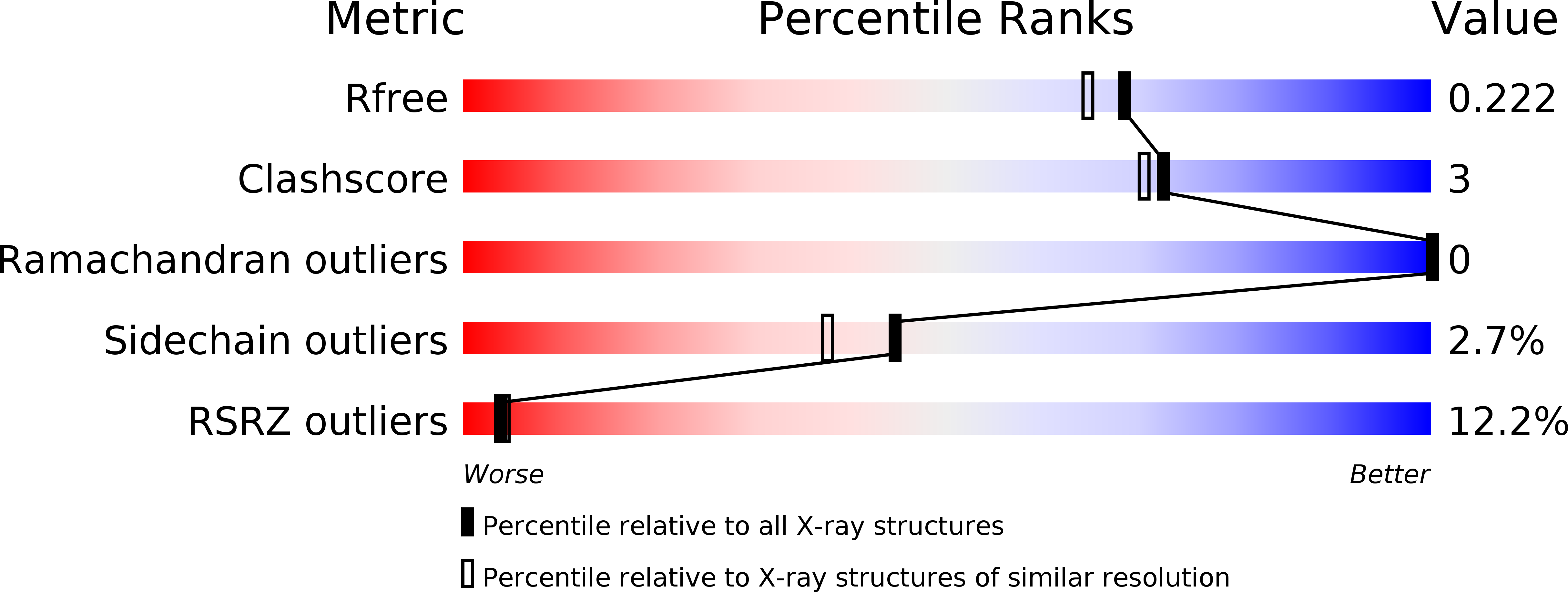
Resolution:
1.90 Å
R-Value Free:
0.22
R-Value Work:
0.17
R-Value Observed:
0.18
Space Group:
P 31 2 1
Macromolecular Entities
Structures with similar UniProt ID
Protein Blast
Polymer Type:polypeptide(L)
Molecule:Protein SflA
Chain IDs:A
Chain Length:133
Number of Molecules:1
Biological Source:Vibrio alginolyticus
Primary Citation
Structure of the periplasmic domain of SflA involved in spatial regulation of the flagellar biogenesis of Vibrio reveals a TPR/SLR-like fold.
J.Biochem. 166 197 204 (2019)
PMID: 30989194 DOI: 10.1093/jb/mvz027

Abstact

Bacteria have evolved various types of flagellum, an organella for bacterial motility, to adapt to their habitat environments. The number and the spatial arrangement of the flagellum are precisely controlled to optimize performance of each type of the flagellar system. Vibrio alginolyticus has a single sheathed flagellum at the cell pole for swimming. SflA is a regulator protein to prevent peritrichous formation of the sheathed flagellum, and consists of an N-terminal periplasmic region, a transmembrane helix, and a C-terminal cytoplasmic region. Whereas the cytoplasmic region has been characterized to be essential for inhibition of the peritrichous growth, the role of the N-terminal region is still unclear. We here determined the structure of the N-terminal periplasmic region of SflA (SflAN) at 1.9-Å resolution. The core of SflAN forms a domain-swapped dimer with tetratricopeptide repeat (TPR)/Sel1-like repeat (SLR) motif, which is often found in the domains responsible for protein-protein interaction in various proteins. The structural similarity and the following mutational analysis based on the structure suggest that SflA binds to unknown partner protein by SflAN and the binding signal is important for the precise control of the SflA function.

Legend

Protein

Chemical

Disease

Primary Citation of related structures