6IEP image
Deposition Date 2018-09-15
Release Date 2019-01-02
Last Version Date 2023-11-22
Entry Detail
PDB ID:
6IEP
Keywords:
Title:
GAPDH of Streptococcus agalactiae
Biological Source:
Source Organism:
Method Details:
Experimental Method:
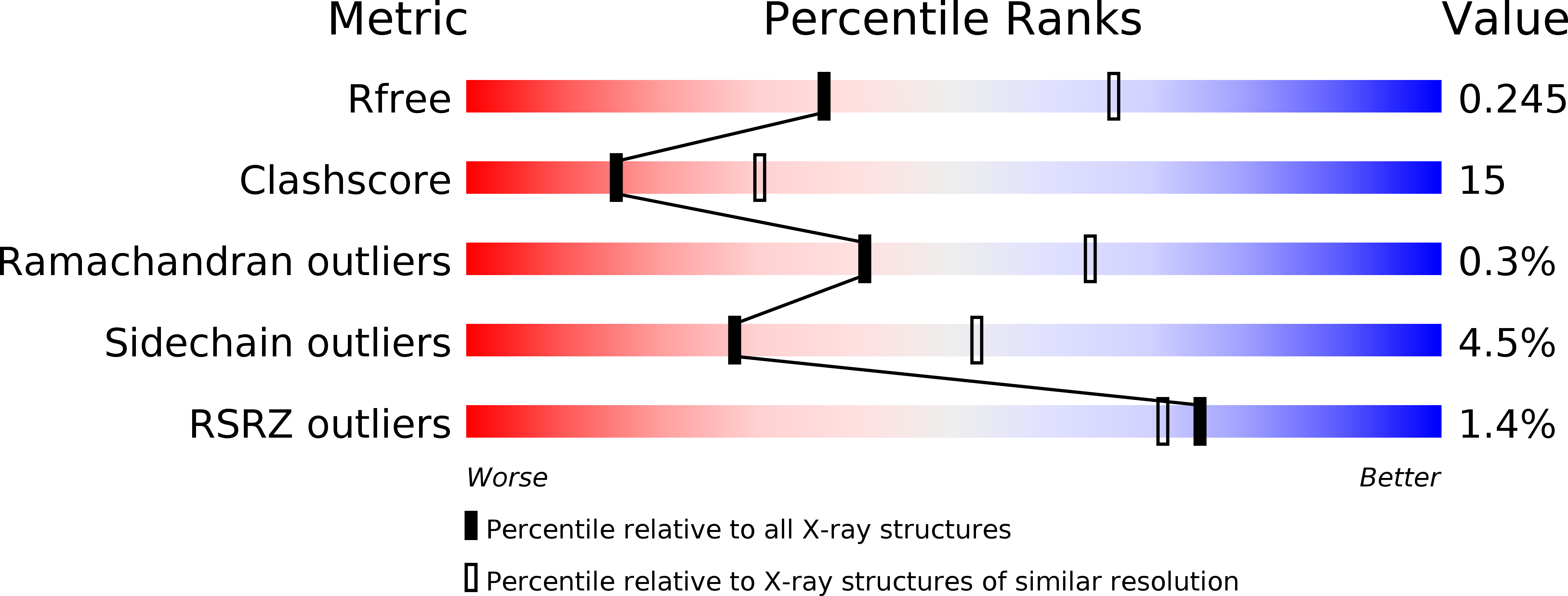
Resolution:
2.60 Å
R-Value Free:
0.24
R-Value Work:
0.20
R-Value Observed:
0.20
Space Group:
P 21 21 21
Macromolecular Entities
Structures with similar UniProt ID
Protein Blast
Polymer Type:polypeptide(L)
Molecule:Glyceraldehyde-3-phosphate dehydrogenase
Gene (Uniprot):gbs1811
Chain IDs:A, B, C, D
Chain Length:344
Number of Molecules:4
Biological Source:Streptococcus agalactiae NEM316
Primary Citation
Crystal structure of GAPDH of Streptococcus agalactiae and characterization of its interaction with extracellular matrix molecules
Microb. Pathog. 127 359 367 (2018)
PMID: 30553015 DOI: 10.1016/j.micpath.2018.12.020

Abstact

GAPDH being a key enzyme in the glycolytic pathway is one of the surface adhesins of many Gram-positive bacteria including Streptococcus agalactiae. This anchorless adhesin is known to bind to host plasminogen (PLG) and fibrinogen (Fg), which enhances the virulence and modulates the host immune system. The crystal structure of the recombinant GAPDH from S. agalactiae (SagGAPDH) was determined at 2.6 Å resolution by molecular replacement. The structure was found to be highly conserved with a typical NAD binding domain and a catalytic domain. In this paper, using biolayer interferometry studies, we report that the multifunctional SagGAPDH enzyme binds to a variety of host molecules such as PLG, Fg, laminin, transferrin and mucin with a KD value of 4.4 × 10-7 M, 9.8 × 10-7 M, 1 × 10-5 M, 9.7 × 10-12 M and 1.4 × 10-7 M respectively. The ligand affinity blots reveal that SagGAPDH binds specifically to α and β subunits of Fg and the competitive binding ELISA assay reveals that the Fg and PLG binding sites on GAPDH does not overlap each other. The PLG binding motif of GAPDH varies with organisms, however positively charged residues in the hydrophobic surroundings is essential for PLG binding. The lysine analogue competitive binding assay and lysine succinylation experiments deciphered the role of SagGAPDH lysines in PLG binding. On structural comparison with S. pneumoniae GAPDH, K171 of SagGAPDH is being predicted to be involved in PLG binding. Further SagGAPDH exhibited enzymatic activity in the presence of Fg, PLG and transferrin. This suggests that these host molecules does not mask the active site and bind at some other region of GAPDH.

Legend

Protein

Chemical

Disease

Primary Citation of related structures