
Deposition Date
2018-11-26
Release Date
2019-11-20
Last Version Date
2024-01-24
Entry Detail
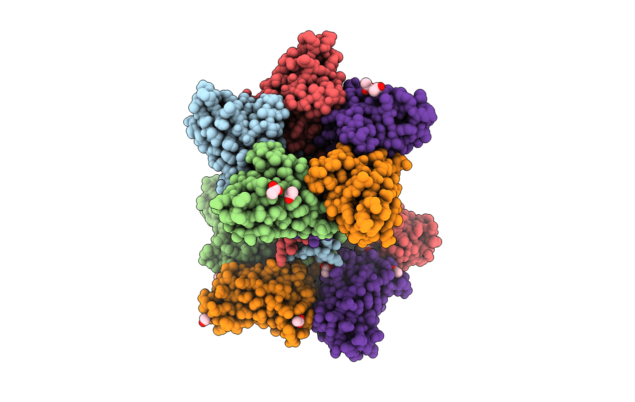
PDB ID:
6IA5
Keywords:
Title:
Crystal Structure Analysis of Bacillus subtilis 168 XepA
Biological Source:
Source Organism(s):
Bacillus subtilis (strain 168) (Taxon ID: 224308)
Expression System(s):
Method Details:
Experimental Method:
Resolution:
1.88 Å
R-Value Free:
0.21
R-Value Work:
0.17
R-Value Observed:
0.17
Space Group:
P 21 21 21
