6I5N image
Deposition Date 2018-11-14
Release Date 2019-05-29
Last Version Date 2025-04-09
Entry Detail
PDB ID:
6I5N
Title:
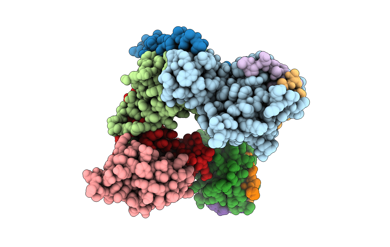
Crystal structure of SOCS2:Elongin C:Elongin B in complex with growth hormone receptor peptide
Biological Source:
Source Organism:
Homo sapiens (Taxon ID: 9606)
Method Details:
Experimental Method:
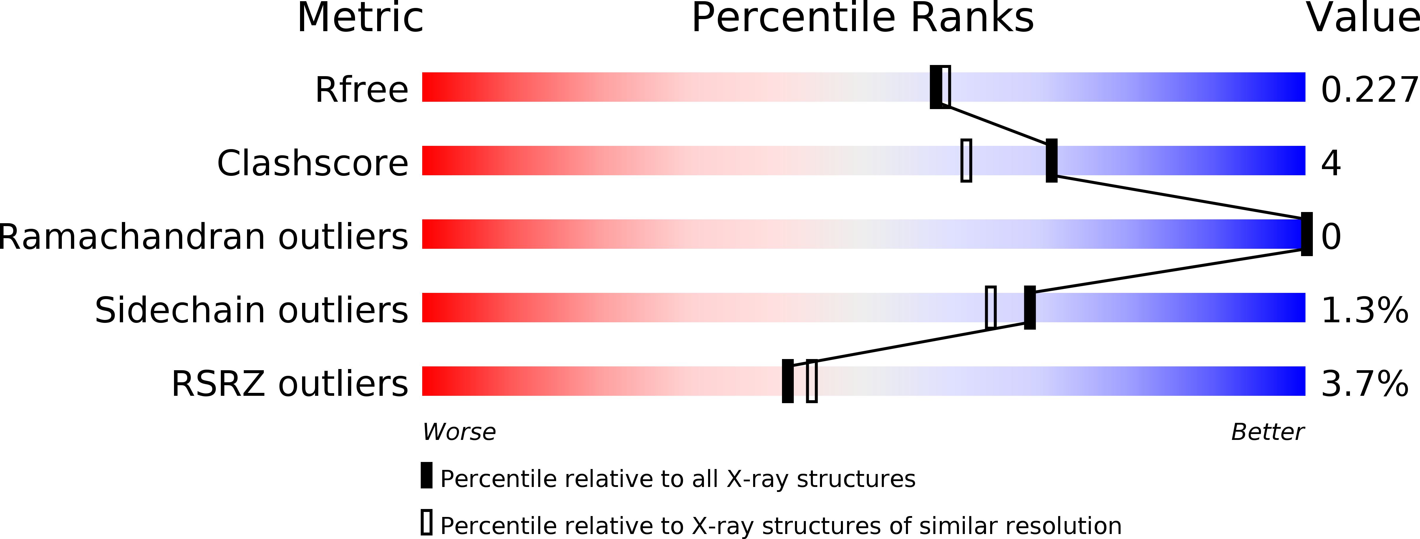
Resolution:
1.98 Å
R-Value Free:
0.22
R-Value Work:
0.18
R-Value Observed:
0.19
Space Group:
P 21 21 2
Macromolecular Entities
Structures with similar UniProt ID
Protein Blast
Polymer Type:polypeptide(L)
Molecule:Suppressor of cytokine signaling 2
Gene (Uniprot):SOCS2
Chain IDs:A, D
Chain Length:169
Number of Molecules:2
Biological Source:Homo sapiens
Structures with similar UniProt ID
Protein Blast
Polymer Type:polypeptide(L)
Molecule:Elongin-B
Gene (Uniprot):ELOB
Chain IDs:B, E
Chain Length:104
Number of Molecules:2
Biological Source:Homo sapiens
Structures with similar UniProt ID
Protein Blast
Polymer Type:polypeptide(L)
Molecule:Elongin-C
Gene (Uniprot):ELOC
Chain IDs:C, F
Chain Length:97
Number of Molecules:2
Biological Source:Homo sapiens
Polymer Type:polypeptide(L)
Molecule:Growth hormone receptor peptide
Chain IDs:G (auth: J), H (auth: K), I, J (auth: L)
Chain Length:11
Number of Molecules:4
Biological Source:Homo sapiens
Modified Residue
Compound ID Chain ID Parent Comp ID Details 2D Image
CAS A CYS modified residue
Primary Citation
Structural insights into substrate recognition by the SOCS2 E3 ubiquitin ligase.
Nat Commun 10 2534 2534 (2019)
PMID: 31182716 DOI: 10.1038/s41467-019-10190-4

Abstact

The suppressor of cytokine signaling 2 (SOCS2) acts as substrate recognition subunit of a Cullin5 E3 ubiquitin ligase complex. SOCS2 binds to phosphotyrosine-modified epitopes as degrons for ubiquitination and proteasomal degradation, yet the molecular basis of substrate recognition has remained elusive. Here, we report co-crystal structures of SOCS2-ElonginB-ElonginC in complex with phosphorylated peptides from substrates growth hormone receptor (GHR-pY595) and erythropoietin receptor (EpoR-pY426) at 1.98 Å and 2.69 Å, respectively. Both peptides bind in an extended conformation recapitulating the canonical SH2 domain-pY pose, but capture different conformations of the EF loop via specific hydrophobic interactions. The flexible BG loop is fully defined in the electron density, and does not contact the substrate degron directly. Cancer-associated SNPs located around the pY pocket weaken substrate-binding affinity in biophysical assays. Our findings reveal insights into substrate recognition and specificity by SOCS2, and provide a blueprint for small molecule ligand design.

Legend

Protein

Chemical

Disease

Primary Citation of related structures
Feedback Form
Name
Email
Institute
Feedback