
Deposition Date
2018-11-13
Release Date
2019-08-14
Last Version Date
2024-01-24
Entry Detail
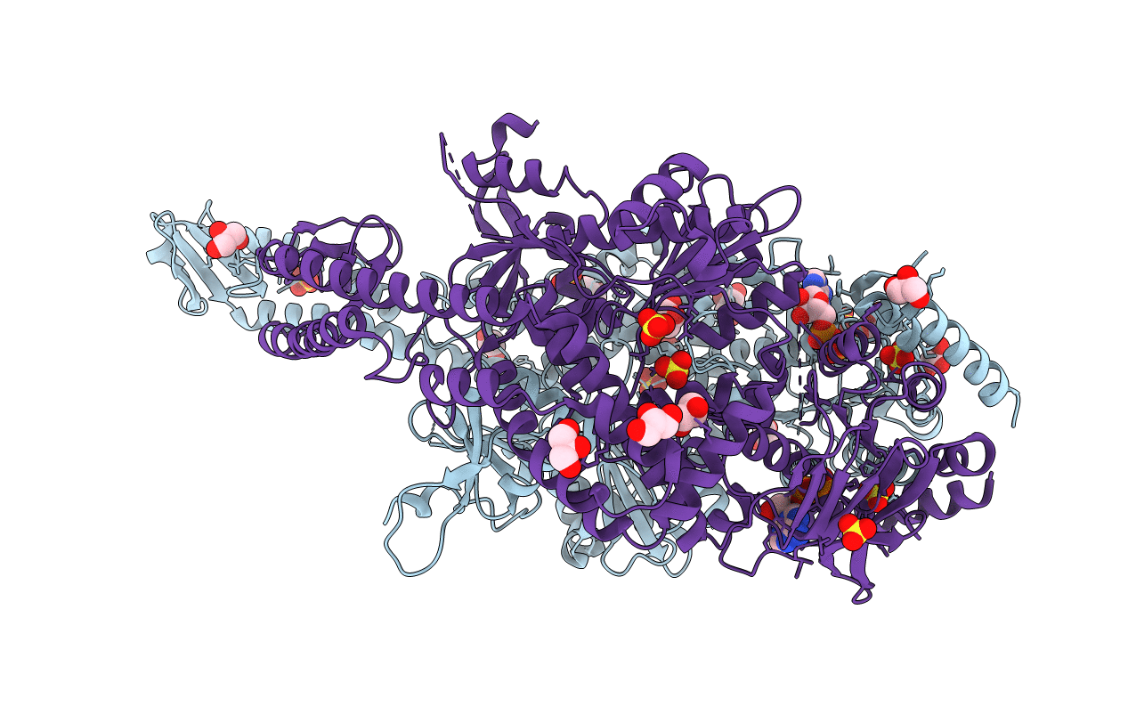
PDB ID:
6I5F
Keywords:
Title:
Crystal structure of DNA-free E.coli MutS P839E dimer mutant
Biological Source:
Source Organism(s):
Escherichia coli (Taxon ID: 562)
Expression System(s):
Method Details:
Experimental Method:
Resolution:
2.60 Å
R-Value Free:
0.25
R-Value Work:
0.20
R-Value Observed:
0.20
Space Group:
P 21 21 21
