Deposition Date
2018-11-13
Release Date
2019-03-13
Last Version Date
2024-05-15
Entry Detail
PDB ID:
6I5C
Keywords:
Title:
Long wavelength native-SAD phasing of Tubulin-Stathmin-TTL complex
Biological Source:
Source Organism(s):
Bos taurus (Taxon ID: 9913)
Rattus norvegicus (Taxon ID: 10116)
Gallus gallus (Taxon ID: 9031)
Rattus norvegicus (Taxon ID: 10116)
Gallus gallus (Taxon ID: 9031)
Expression System(s):
Method Details:
Experimental Method:
Resolution:
2.95 Å
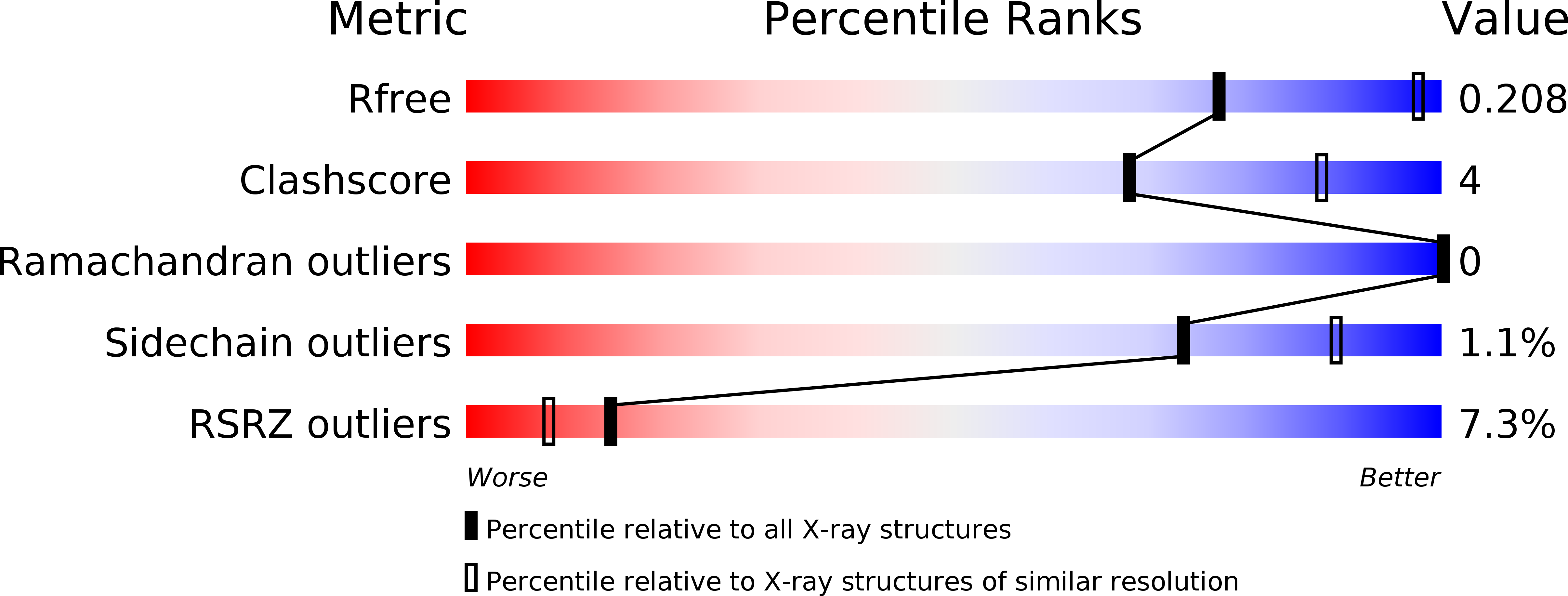
R-Value Free:
0.20
R-Value Work:
0.16
R-Value Observed:
0.17
Space Group:
P 21 21 21