
Deposition Date
2018-11-07
Release Date
2019-09-11
Last Version Date
2024-01-24
Entry Detail
PDB ID:
6I3T
Keywords:
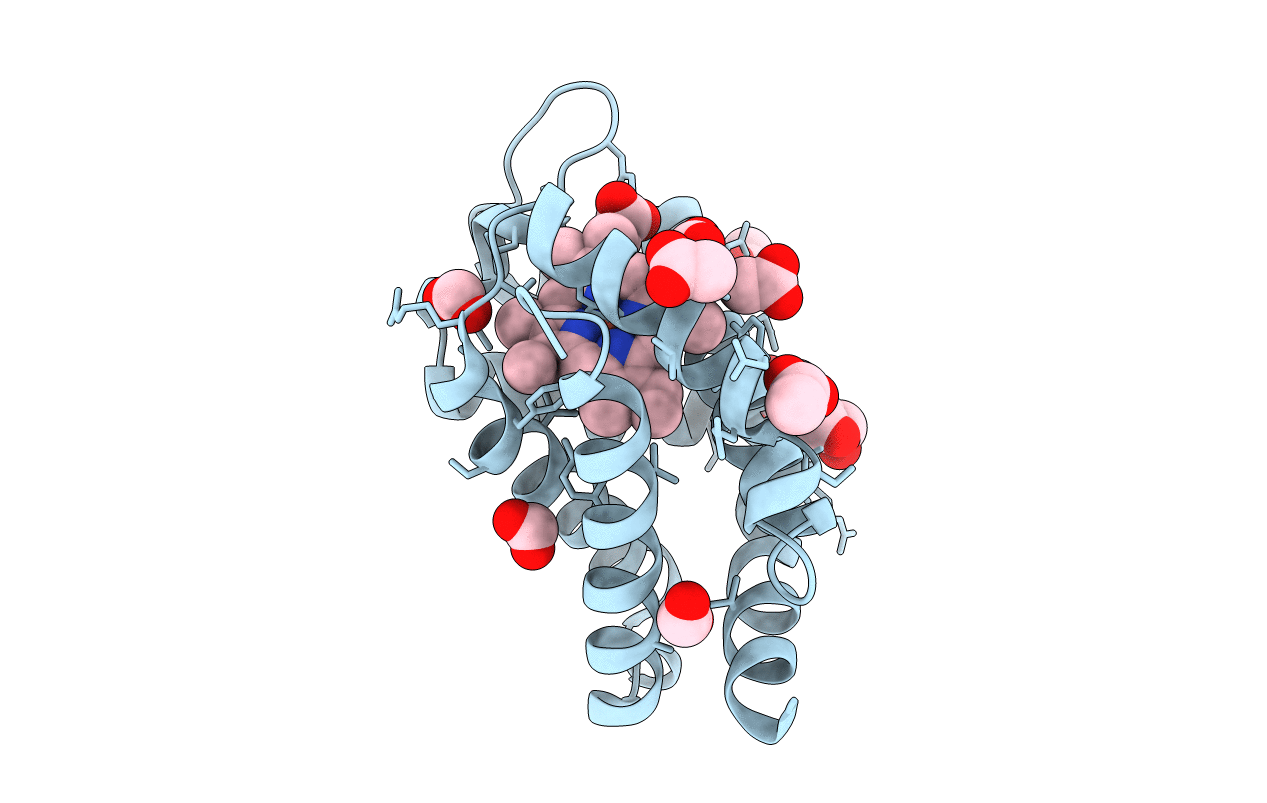
Title:
Crystal structure of murine neuroglobin bound to CO at 40 K.
Biological Source:
Source Organism(s):
Mus musculus (Taxon ID: 10090)
Expression System(s):
Method Details:
Experimental Method:
Resolution:
2.00 Å
R-Value Free:
0.22
R-Value Work:
0.16
R-Value Observed:
0.16
Space Group:
H 3 2
