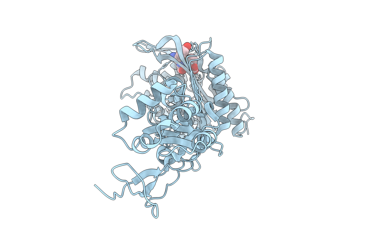
6I1E image
Entry Detail
PDB ID:
6I1E
Title:
Crystal structure of Pseudomonas aeruginosa Penicillin-Binding Protein 3 in complex with amoxicillin
Biological Source:
Source Organism:
PDB Version:
Deposition Date:
2018-10-28
Release Date:
2019-11-20
Method Details:
Experimental Method:
Resolution:
1.64 Å
R-Value Free:
0.23
R-Value Work:
0.21
R-Value Observed:
0.21
Space Group:
P 21 21 21
Macromolecular Entities
Polymer Type:polypeptide(L)
Description:Peptidoglycan D,D-transpeptidase FtsI
Chain IDs:A
Chain Length:537
Number of Molecules:1
Biological Source:Pseudomonas aeruginosa
Ligand Molecules
Primary Citation
Novel and Improved Crystal Structures of H. influenzae, E. coli and P. aeruginosa Penicillin-Binding Protein 3 (PBP3) and N. gonorrhoeae PBP2: Toward a Better Understanding of beta-Lactam Target-Mediated Resistance.
J.Mol.Biol. 431 3501 3519 (2019)
PMID: 31301409 DOI: 10.1016/j.jmb.2019.07.010

Abstact

Even with the emergence of antibiotic resistance, penicillin and the wider family of β-lactams have remained the single most important family of antibiotics. The periplasmic/extra-cytoplasmic targets of penicillin are a family of enzymes with a highly conserved catalytic activity involved in the final stage of bacterial cell wall (peptidoglycan) biosynthesis. Named after their ability to bind penicillin, rather than their catalytic activity, these key targets are called penicillin-binding proteins (PBPs). Resistance is predominantly mediated by reducing the target drug concentration via β-lactamases; however, naturally transformable bacteria have also acquired target-mediated resistance by inter-species recombination. Here we focus on structural based interpretations of amino acid alterations associated with the emergence of resistance within clinical isolates and include new PBP3 structures along with new, and improved, PBP-β-lactam co-structures.

Legend

Protein

Chemical

Disease

Primary Citation of related structures