Deposition Date
2018-10-28
Release Date
2020-01-29
Last Version Date
2024-11-13
Entry Detail
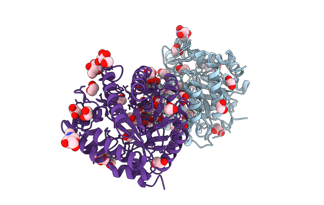
Biological Source:
Source Organism(s):
Aspergillus niger (Taxon ID: 5061)
Expression System(s):
Method Details:
Experimental Method:
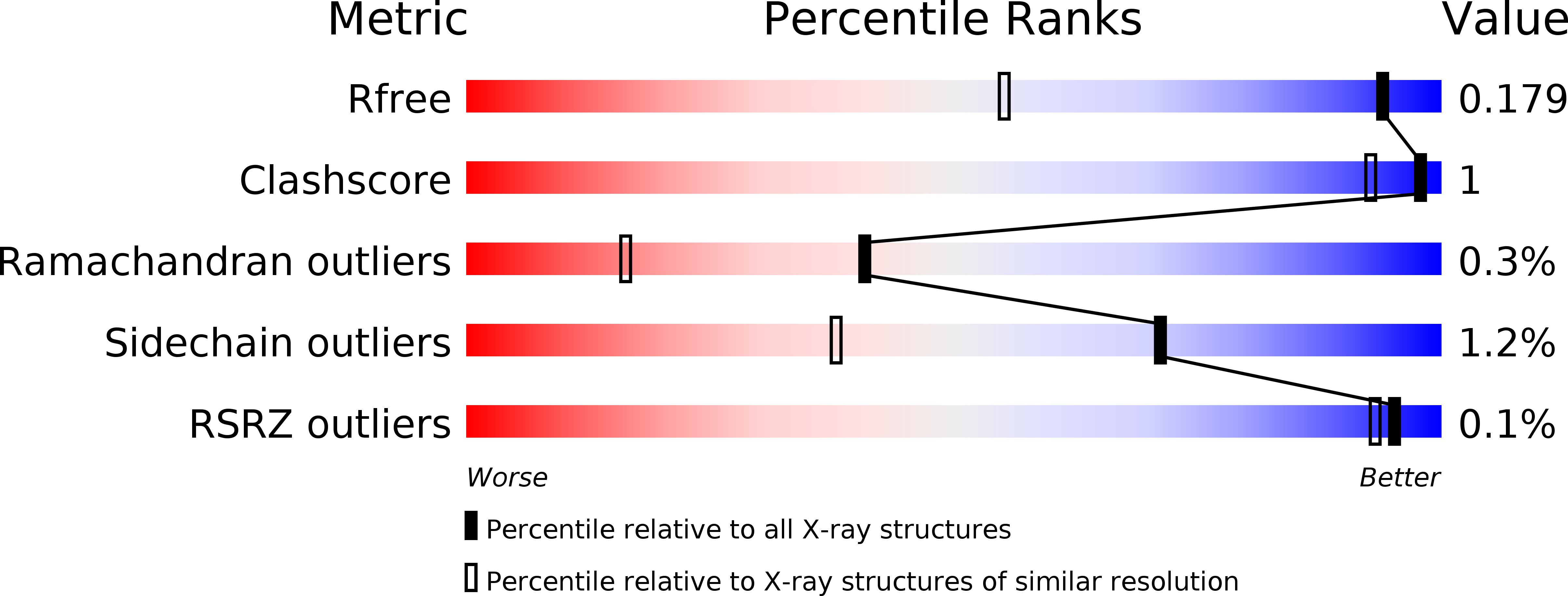
Resolution:
1.27 Å
R-Value Free:
0.17
R-Value Work:
0.15
R-Value Observed:
0.15
Space Group:
P 21 21 21