6I19 image
Deposition Date 2018-10-27
Release Date 2018-12-05
Last Version Date 2024-10-09
Entry Detail
PDB ID:
6I19
Title:
Crystal structure of Chlamydomonas reinhardtii thioredoxin h1
Biological Source:
Source Organism:
Host Organism:
Method Details:
Experimental Method:
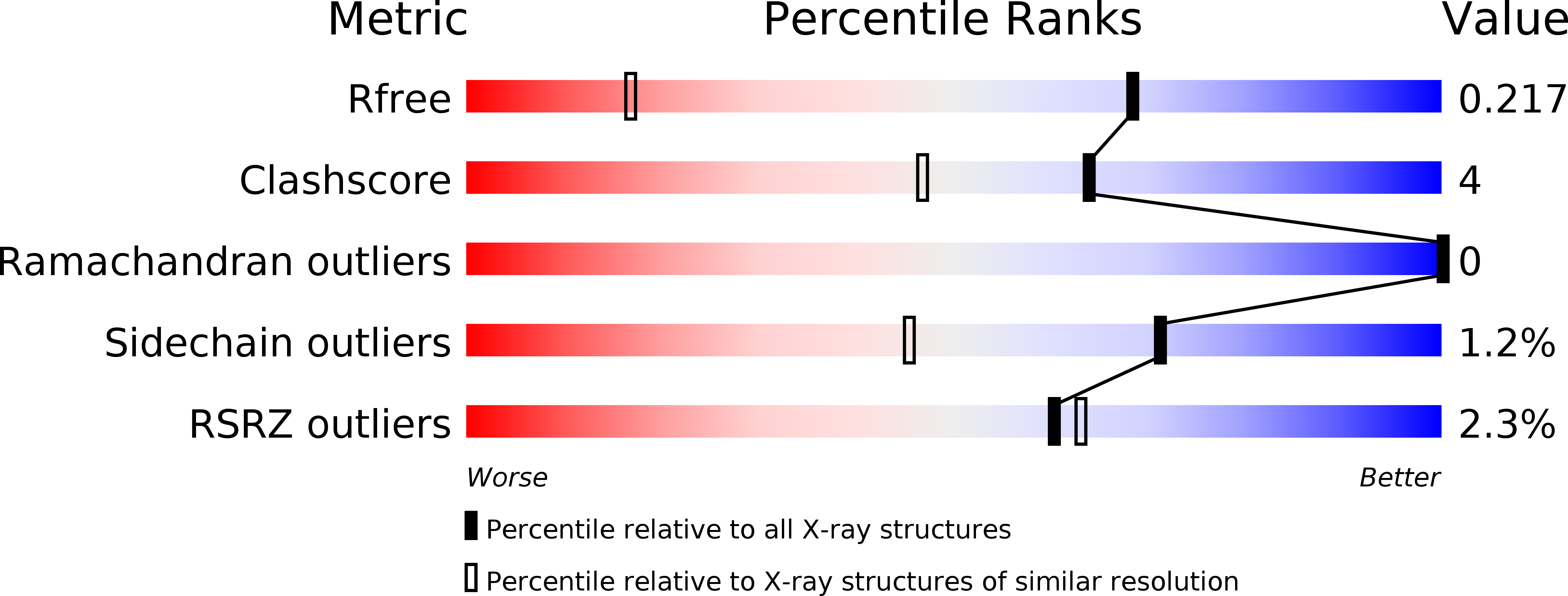
Resolution:
1.38 Å
R-Value Free:
0.21
R-Value Work:
0.18
R-Value Observed:
0.18
Space Group:
P 31 2 1
Macromolecular Entities
Structures with similar UniProt ID
Protein Blast
Polymer Type:polypeptide(L)
Molecule:Thioredoxin H-type
Gene (Uniprot):TRXH
Chain IDs:A, B
Chain Length:113
Number of Molecules:2
Biological Source:Chlamydomonas reinhardtii
Primary Citation
Crystal Structure of Chloroplastic Thioredoxin f2 fromChlamydomonas reinhardtiiReveals Distinct Surface Properties.
Antioxidants (Basel) 7 ? ? (2018)
PMID: 30477165 DOI: 10.3390/antiox7120171

Abstact

Protein disulfide reduction by thioredoxins (TRXs) controls the conformation of enzyme active sites and their multimeric complex formation. TRXs are small oxidoreductases that are broadly conserved in all living organisms. In photosynthetic eukaryotes, TRXs form a large multigenic family, and they have been classified in different types: f, m, x, y, and z types are chloroplastic, while o and h types are located in mitochondria and cytosol. In the model unicellular alga Chlamydomonas reinhardtii, the TRX family contains seven types, with f- and h-types represented by two isozymes. Type-f TRXs interact specifically with targets in the chloroplast, controlling photosynthetic carbon fixation by the Calvin⁻Benson cycle. We solved the crystal structures of TRX f2 and TRX h1 from C. reinhardtii. The systematic comparison of their atomic features revealed a specific conserved electropositive crown around the active site of TRX f, complementary to the electronegative surface of their targets. We postulate that this surface provides specificity to each type of TRX.

Legend

Protein

Chemical

Disease

Primary Citation of related structures