6HZK image
Deposition Date 2018-10-23
Release Date 2019-03-27
Last Version Date 2024-10-09
Entry Detail
PDB ID:
6HZK
Keywords:
Title:
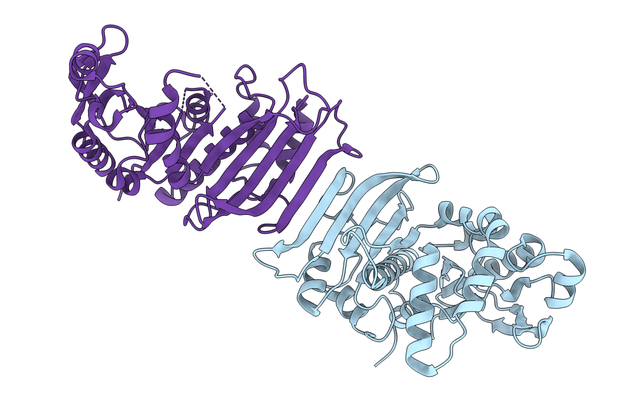
Crystal structure of redox-inhibited phosphoribulokinase from Synechococcus sp. (strain PCC 6301)
Biological Source:
Method Details:
Experimental Method:
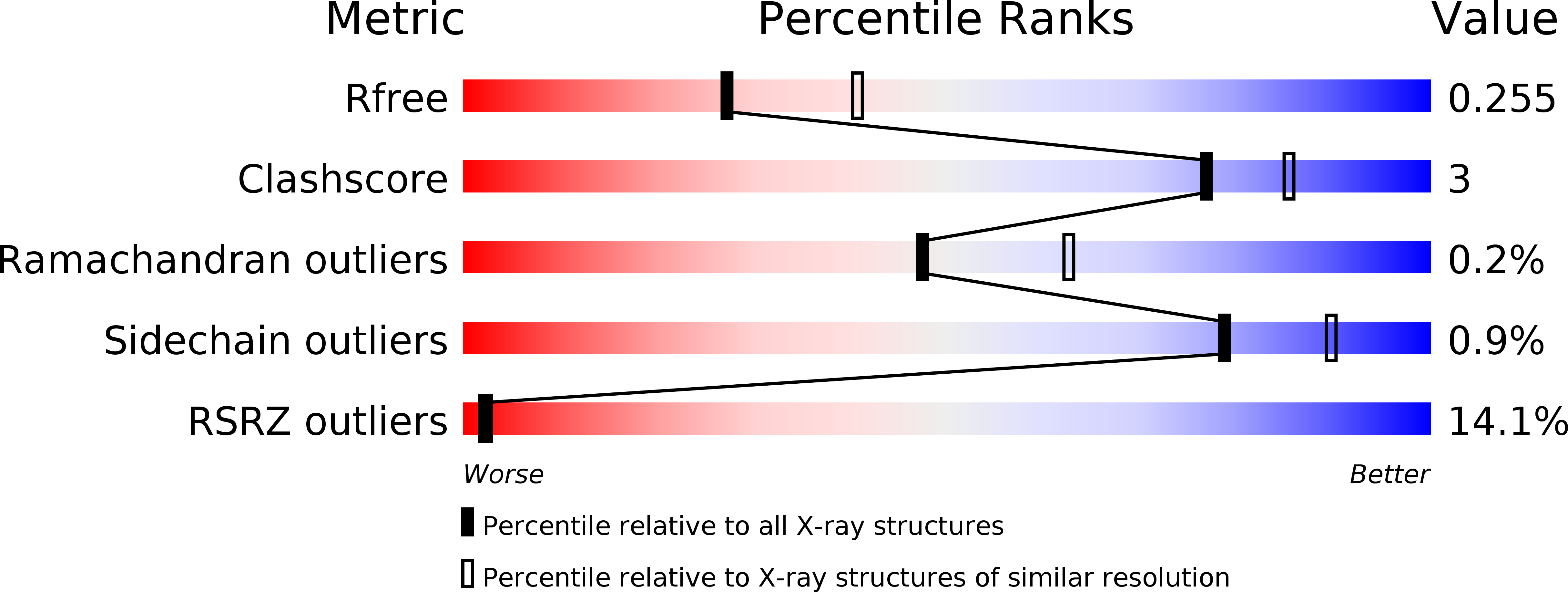
Resolution:
2.40 Å
R-Value Free:
0.25
R-Value Work:
0.22
R-Value Observed:
0.22
Space Group:
H 3 2
Macromolecular Entities
Polymer Type:polypeptide(L)
Molecule:Phosphoribulokinase
Gene (Uniprot):prk
Chain IDs:A, B
Chain Length:333
Number of Molecules:2
Biological Source:Synechococcus sp. (strain ATCC 27144 / PCC 6301 / SAUG 1402/1)
Primary Citation
Crystal structure of phosphoribulokinase from Synechococcus sp. strain PCC 6301.
Acta Crystallogr.,Sect.F 75 278 289 (2019)
PMID: 30950829 DOI: 10.1107/S2053230X19002693

Abstact

Phosphoribulokinase (PRK) catalyses the ATP-dependent phosphorylation of ribulose 5-phosphate to give ribulose 1,5-bisphosphate. Regulation of this reaction in response to light controls carbon fixation during photosynthesis. Here, the crystal structure of PRK from the cyanobacterium Synechococcus sp. strain PCC 6301 is presented. The enzyme is dimeric and has an α/β-fold with an 18-stranded β-sheet at its core. Interestingly, a disulfide bond is found between Cys40 and the P-loop residue Cys18, revealing the structural basis for the redox inactivation of PRK activity. A second disulfide bond appears to rigidify the dimer interface and may thereby contribute to regulation by the adaptor protein CP12 and glyceraldehyde-3-phosphate dehydrogenase.

Legend

Protein

Chemical

Disease

Primary Citation of related structures