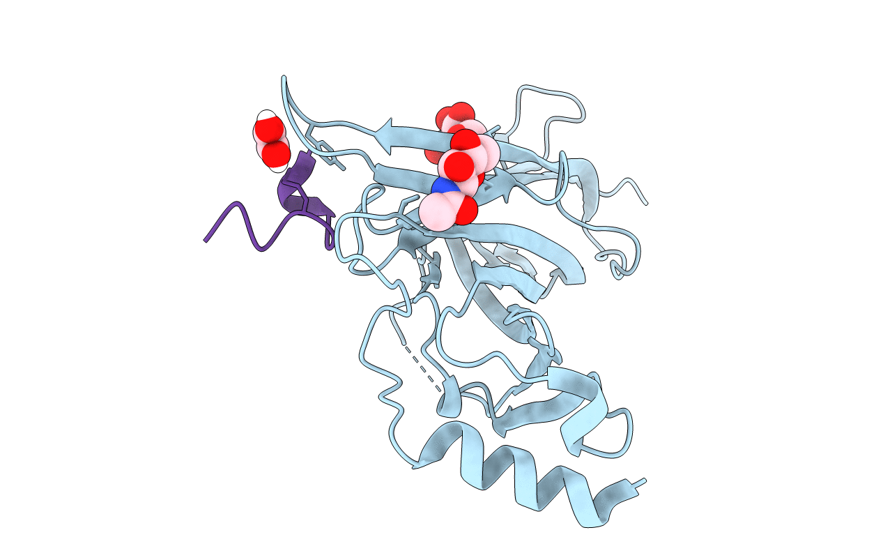
6HY7 image
Entry Detail
PDB ID:
6HY7
Keywords:
Title:
Crystal structure of alpha9 nAChR extracellular domain in complex with alpha-conotoxin RgIA
Biological Source:
Source Organism:
Host Organism:
PDB Version:
Deposition Date:
2018-10-19
Release Date:
2019-05-22
Method Details:
Experimental Method:
Resolution:
2.26 Å
R-Value Free:
0.24
R-Value Work:
0.19
R-Value Observed:
0.19
Space Group:
P 21 21 2
Macromolecular Entities
Polymer Type:polypeptide(L)
Description:Neuronal acetylcholine receptor subunit alpha-9
Chain IDs:A
Chain Length:218
Number of Molecules:1
Biological Source:Homo sapiens
Polymer Type:polypeptide(L)
Description:Alpha-conotoxin RgIA
Chain IDs:B
Chain Length:13
Number of Molecules:1
Biological Source:Conus regius
Modified Residue
Compound ID Chain ID Parent Comp ID Details 2D Image
AAR B ARG modified residue
Primary Citation
Crystal Structure of the Monomeric Extracellular Domain of alpha 9 Nicotinic Receptor Subunit in Complex With alpha-Conotoxin RgIA: Molecular Dynamics Insights Into RgIA Binding to alpha 9 alpha 10 Nicotinic Receptors.
Front Pharmacol 10 474 474 (2019)
PMID: 31118896 DOI: 10.3389/fphar.2019.00474

Abstact

The α9 subunit of nicotinic acetylcholine receptors (nAChRs) exists mainly in heteropentameric assemblies with α10. Accumulating data indicate the presence of three different binding sites in α9α10 nAChRs: the α9(+)/α9(-), the α9(+)/α10(-), and the α10(+)/α9(-). The major role of the principal (+) side of the extracellular domain (ECD) of α9 subunit in binding of the antagonists methyllylcaconitine and α-bungarotoxin was shown previously by the crystal structures of the monomeric α9-ECD with these molecules. Here we present the 2.26-Å resolution crystal structure of α9-ECD in complex with α-conotoxin (α-Ctx) RgIA, a potential drug for chronic pain, the first structure reported for a complex between an nAChR domain and an α-Ctx. Superposition of this structure with those of other α-Ctxs bound to the homologous pentameric acetylcholine binding proteins revealed significant similarities in the orientation of bound conotoxins, despite the monomeric state of the α9-ECD. In addition, ligand-binding studies calculated a binding affinity of RgIA to the α9-ECD at the low micromolar range. Given the high identity between α9 and α10 ECDs, particularly at their (+) sides, the presented structure was used as template for molecular dynamics simulations of the ECDs of the human α9α10 nAChR in pentameric assemblies. Our results support a favorable binding of RgIA at α9(+)/α9(-) or α10(+)/α9(-) rather than the α9(+)/α10(-) interface, in accordance with previous mutational and functional data.

Legend

Protein

Chemical

Disease

Primary Citation of related structures