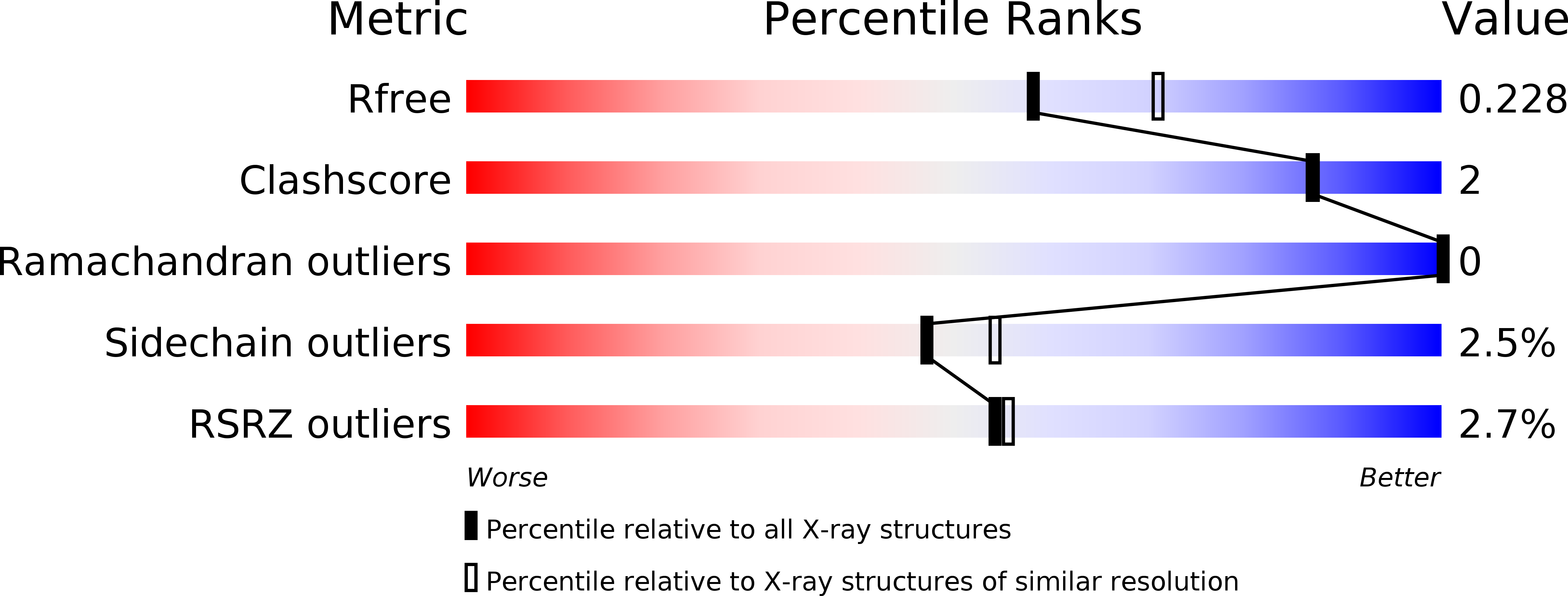
6HVN image
Deposition Date 2018-10-11
Release Date 2019-06-05
Last Version Date 2024-05-15
Entry Detail
PDB ID:
6HVN
Keywords:
Title:
CdaA-APO Y187A Mutant
Biological Source:
Method Details:
Experimental Method:
Resolution:
2.23 Å
R-Value Free:
0.22
R-Value Work:
0.18
R-Value Observed:
0.18
Space Group:
P 21 21 21
Macromolecular Entities
Structures with similar UniProt ID
Protein Blast
Polymer Type:polypeptide(L)
Molecule:Diadenylate cyclase
Gene (Uniprot):dacA
Mutagens:Y187A
Chain IDs:A, B
Chain Length:178
Number of Molecules:2
Biological Source:Listeria monocytogenes serovar 1/2a (strain ATCC BAA-679 / EGD-e)
Ligand Molecules
Peptide-like Molecules
PRD_900003
Primary Citation
Crystal structures of the c-di-AMP-synthesizing enzyme CdaA.
J.Biol.Chem. 294 10463 10470 (2019)
PMID: 31118276 DOI: 10.1074/jbc.RA119.009246

Abstact

Cyclic di-AMP (c-di-AMP) is the only second messenger known to be essential for bacterial growth. It has been found mainly in Gram-positive bacteria, including pathogenic bacteria like Listeria monocytogenes CdaA is the sole diadenylate cyclase in L. monocytogenes, making this enzyme an attractive target for the development of novel antibiotic compounds. Here we report crystal structures of CdaA from L. monocytogenes in the apo state, in the post-catalytic state with bound c-di-AMP and catalytic Co2+ ions, as well as in a complex with AMP. These structures reveal the flexibility of a tyrosine side chain involved in locking the adenine ring after ATP binding. The essential role of this tyrosine was confirmed by mutation to Ala, leading to drastic loss of enzymatic activity.

Legend

Protein

Chemical

Disease

Primary Citation of related structures