Deposition Date
2018-10-05
Release Date
2019-01-23
Last Version Date
2024-11-13
Entry Detail
PDB ID:
6HU5
Keywords:
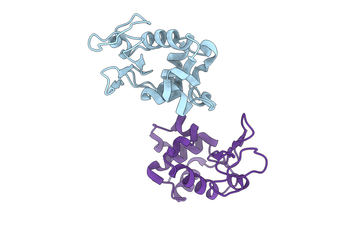
Title:
STRUCTURE OF HEWL BY ELECTRON DIFFRACTION AND MICROFOCUS DIFFRACTION
Biological Source:
Source Organism(s):
Gallus gallus (Taxon ID: 9031)
Method Details:
Experimental Method:
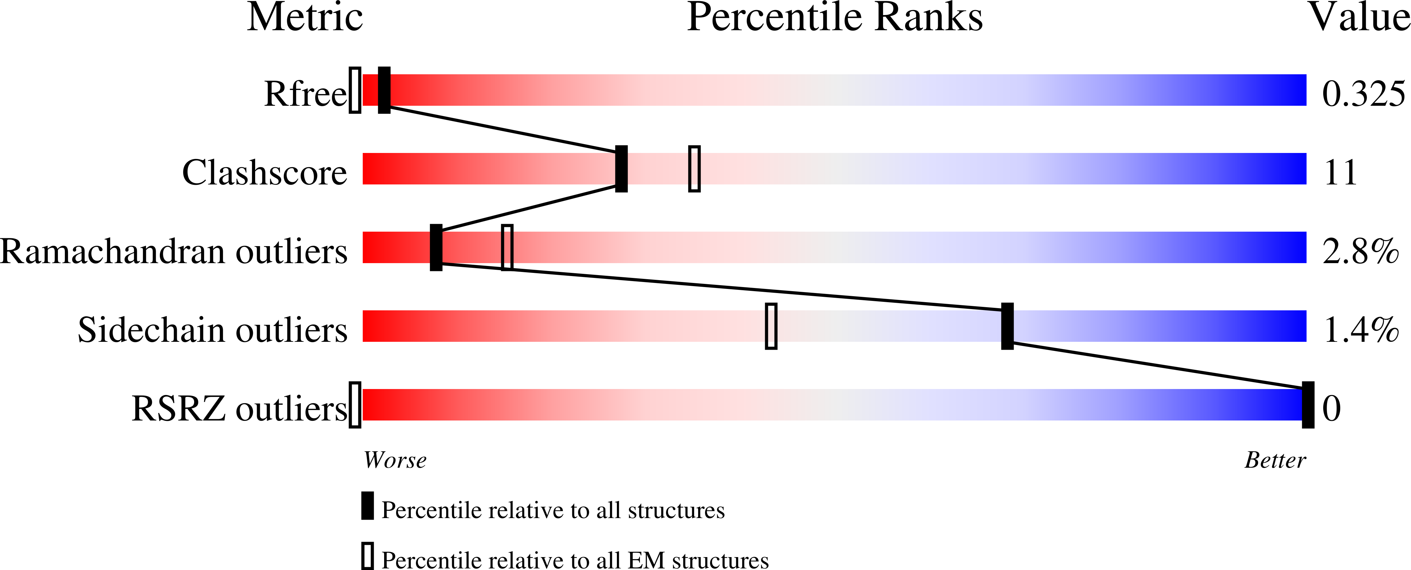
Resolution:
2.80 Å
R-Value Free:
0.33
R-Value Work:
0.29
R-Value Observed:
0.29
Space Group:
P 1 21 1