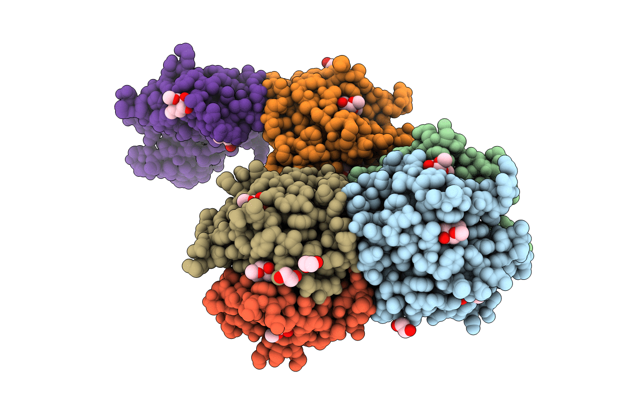
6HTN image
Deposition Date 2018-10-04
Release Date 2019-01-23
Last Version Date 2024-05-01
Entry Detail
PDB ID:
6HTN
Title:
Structure of a fucose lectin from Kordia zhangzhouensis in complex with methyl-fucoside
Biological Source:
Source Organism:
Host Organism:
Method Details:
Experimental Method:
Resolution:
1.55 Å
R-Value Free:
0.18
R-Value Work:
0.15
R-Value Observed:
0.15
Space Group:
P 2 21 21
Macromolecular Entities
Polymer Type:polypeptide(L)
Molecule:Fucose-binding lectin
Gene (Uniprot):C8N46_102337
Chain IDs:A, B, C, D, E, F
Chain Length:144
Number of Molecules:6
Biological Source:Kordia periserrulae
Primary Citation
Architecture and Evolution of Blade Assembly in beta-propeller Lectins.
Structure 27 764 775.e3 (2019)
PMID: 30853410 DOI: 10.1016/j.str.2019.02.002

Abstact

Lectins with a β-propeller fold bind glycans on the cell surface through multivalent binding sites and appropriate directionality. These proteins are formed by repeats of short domains, raising questions about evolutionary duplication. However, these repeats are difficult to detect in translated genomes and seldom correctly annotated in sequence databases. To address these issues, we defined the blade signature of the five types of β-propellers using 3D-structural data. With these templates, we predicted 3,887 β-propeller lectins in 1,889 species and organized this information in a searchable online database. The data reveal a widespread distribution of β-propeller lectins across species. Prediction also emphasizes multiple architectures and led to the discovery of a β-propeller assembly scenario. This was confirmed by producing and characterizing a predicted protein coded in the genome of Kordia zhangzhouensis. The crystal structure uncovers an intermediate in the evolution of β-propeller assembly and demonstrates the power of our tools.

Legend

Protein

Chemical

Disease

Primary Citation of related structures