6HSW image
Deposition Date 2018-10-02
Release Date 2019-03-20
Last Version Date 2024-11-13
Entry Detail
PDB ID:
6HSW
Keywords:
Title:
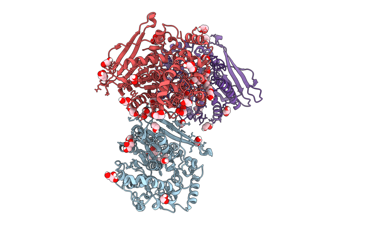
A CE15 glucuronoyl esterase from Teredinibacter turnerae T7901
Biological Source:
Source Organism:
Method Details:
Experimental Method:
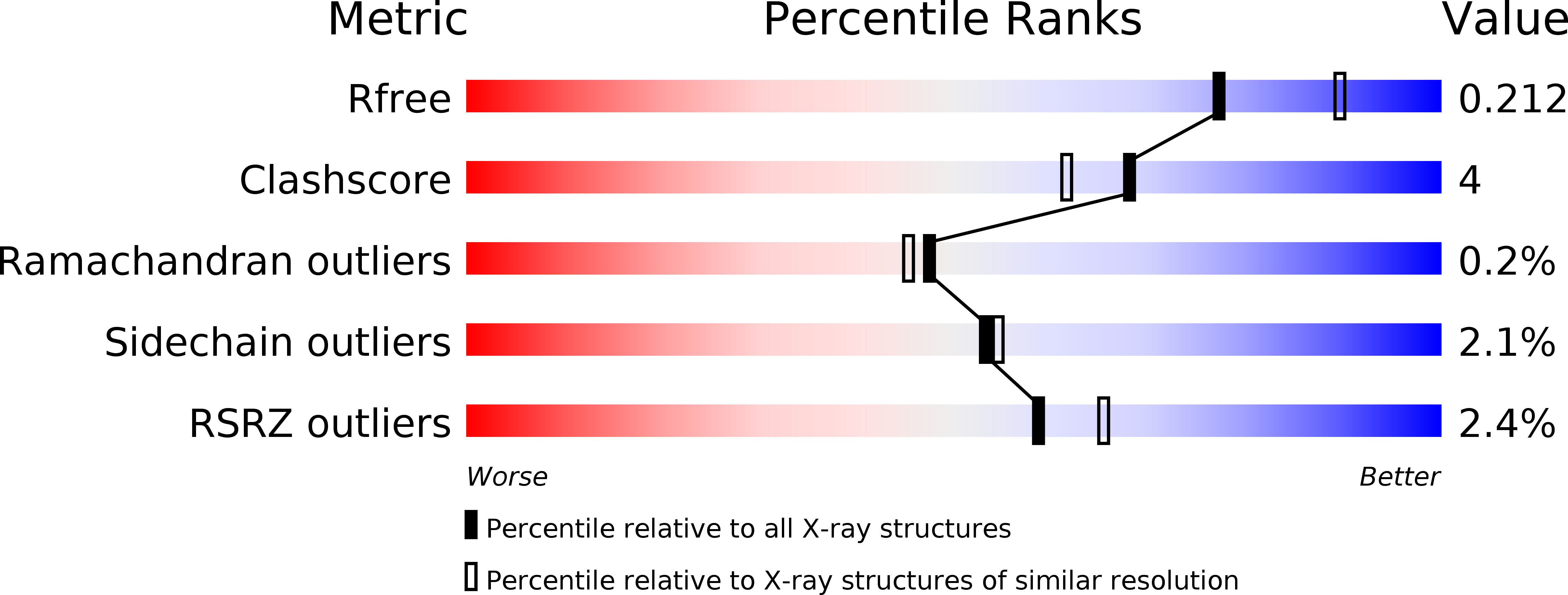
Resolution:
2.15 Å
R-Value Free:
0.21
R-Value Work:
0.16
R-Value Observed:
0.16
Space Group:
P 31 2 1
Macromolecular Entities
Structures with similar UniProt ID
Protein Blast
Polymer Type:polypeptide(L)
Molecule:Carbohydrate esterase family 15 domain protein
Gene (Uniprot):TERTU_0517
Chain IDs:A, B, C
Chain Length:440
Number of Molecules:3
Biological Source:Teredinibacter turnerae T7901
Modified Residue
Compound ID Chain ID Parent Comp ID Details 2D Image
MSE A ? modified residue
Primary Citation
Structure-function analyses reveal that a glucuronoyl esterase fromTeredinibacter turneraeinteracts with carbohydrates and aromatic compounds.
J.Biol.Chem. 294 6635 6644 (2019)
PMID: 30814248 DOI: 10.1074/jbc.RA119.007831

Abstact

Glucuronoyl esterases (GEs) catalyze the cleavage of ester linkages found between lignin and glucuronic acid moieties on glucuronoxylan in plant biomass. As such, GEs represent promising biochemical tools in industrial processing of these recalcitrant resources. However, details on how GEs interact with their natural substrates are sparse, calling for thorough structure-function studies. Presented here is the structure and biochemical characterization of a GE, TtCE15A, from the bacterium Teredinibacter turnerae, a symbiont of wood-boring shipworms. To gain deeper insight into enzyme-substrate interactions, inhibition studies were performed with both the WT TtCE15A and variants in which we, by using site-directed mutagenesis, substituted residues suggested to have key roles in binding to or interacting with the aromatic and carbohydrate structures of its uronic acid ester substrates. Our results support the hypothesis that two aromatic residues (Phe-174 and Trp-376), conserved in bacterial GEs, interact with aromatic and carbohydrate structures of these substrates in the enzyme active site, respectively. The solved crystal structure of TtCE15A revealed features previously not observed in either fungal or bacterial GEs, with a large inserted N-terminal region neighboring the active site and a differently positioned residue of the catalytic triad. The findings highlight key interactions between GEs and complex lignin-carbohydrate ester substrates and advance our understanding of the substrate specificities of these enzymes in biomass conversion.

Legend

Protein

Chemical

Disease

Primary Citation of related structures