
Deposition Date
2018-10-01
Release Date
2019-01-30
Last Version Date
2024-05-01
Entry Detail
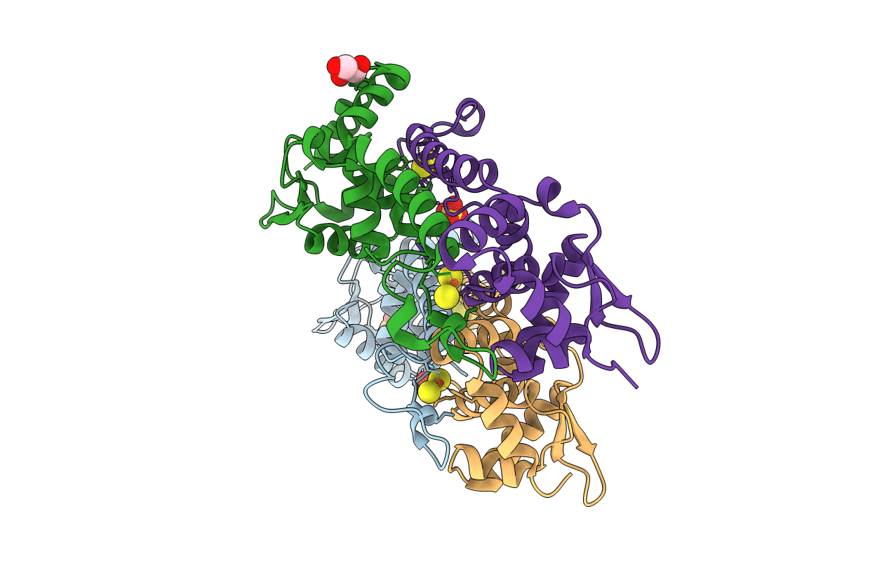
PDB ID:
6HSE
Keywords:
Title:
Structure of dithionite-reduced RsrR in spacegroup P2(1)
Biological Source:
Source Organism(s):
Expression System(s):
Method Details:
Experimental Method:
Resolution:
2.30 Å
R-Value Free:
0.26
R-Value Work:
0.22
R-Value Observed:
0.23
Space Group:
P 1 21 1
