6HRV image
Deposition Date 2018-09-28
Release Date 2019-01-02
Last Version Date 2024-11-06
Entry Detail
PDB ID:
6HRV
Keywords:
Title:
Crystal structure of the zebrafish peroxisomal SCP2-thiolase (type-1)
Biological Source:
Source Organism:
Danio rerio (Taxon ID: 7955)
Method Details:
Experimental Method:
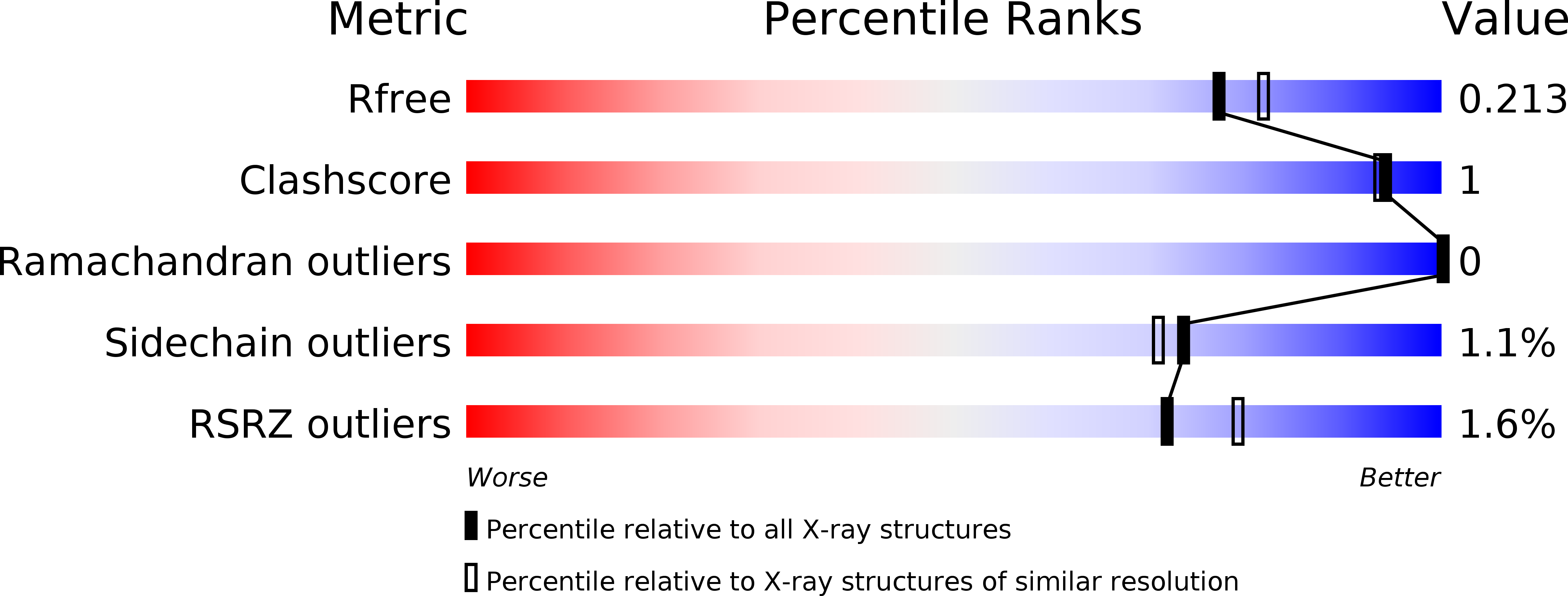
Resolution:
1.95 Å
R-Value Free:
0.20
R-Value Work:
0.16
R-Value Observed:
0.17
Space Group:
C 1 2 1
Macromolecular Entities
Polymer Type:polypeptide(L)
Molecule:SCP2-thiolase (type-1)
Gene (Uniprot):scp2a
Chain IDs:A
Chain Length:421
Number of Molecules:1
Biological Source:Danio rerio
Polymer Type:polypeptide(L)
Molecule:SCP2-thiolase (type-1)
Gene (Uniprot):scp2a
Chain IDs:B
Chain Length:421
Number of Molecules:1
Biological Source:Danio rerio
Modified Residue
Compound ID Chain ID Parent Comp ID Details 2D Image
CSO B CYS modified residue
Primary Citation
The peroxisomal zebrafish SCP2-thiolase (type-1) is a weak transient dimer as revealed by crystal structures and native mass spectrometry.
Biochem. J. 476 307 332 (2019)
PMID: 30573650 DOI: 10.1042/BCJ20180788

Abstact

The SCP2 (sterol carrier protein 2)-thiolase (type-1) functions in the vertebrate peroxisomal, bile acid synthesis pathway, converting 24-keto-THC-CoA and CoA into choloyl-CoA and propionyl-CoA. This conversion concerns the β-oxidation chain shortening of the steroid fatty acyl-moiety of 24-keto-THC-CoA. This class of dimeric thiolases has previously been poorly characterized. High-resolution crystal structures of the zebrafish SCP2-thiolase (type-1) now reveal an open catalytic site, shaped by residues of both subunits. The structure of its non-dimerized monomeric form has also been captured in the obtained crystals. Four loops at the dimer interface adopt very different conformations in the monomeric form. These loops also shape the active site and their structural changes explain why a competent active site is not present in the monomeric form. Native mass spectrometry studies confirm that the zebrafish SCP2-thiolase (type-1) as well as its human homolog are weak transient dimers in solution. The crystallographic binding studies reveal the mode of binding of CoA and octanoyl-CoA in the active site, highlighting the conserved geometry of the nucleophilic cysteine, the catalytic acid/base cysteine and the two oxyanion holes. The dimer interface of SCP2-thiolase (type-1) is equally extensive as in other thiolase dimers; however, it is more polar than any of the corresponding interfaces, which correlates with the notion that the enzyme forms a weak transient dimer. The structure comparison of the monomeric and dimeric forms suggests functional relevance of this property. These comparisons provide also insights into the structural rearrangements that occur when the folded inactive monomers assemble into the mature dimer.

Legend

Protein

Chemical

Disease

Primary Citation of related structures