6HPB image
Deposition Date 2018-09-20
Release Date 2019-09-18
Last Version Date 2024-10-16
Entry Detail
PDB ID:
6HPB
Keywords:
Title:
Crystal structure of the E.coli HicAB toxin-antitoxin complex
Biological Source:
Method Details:
Experimental Method:
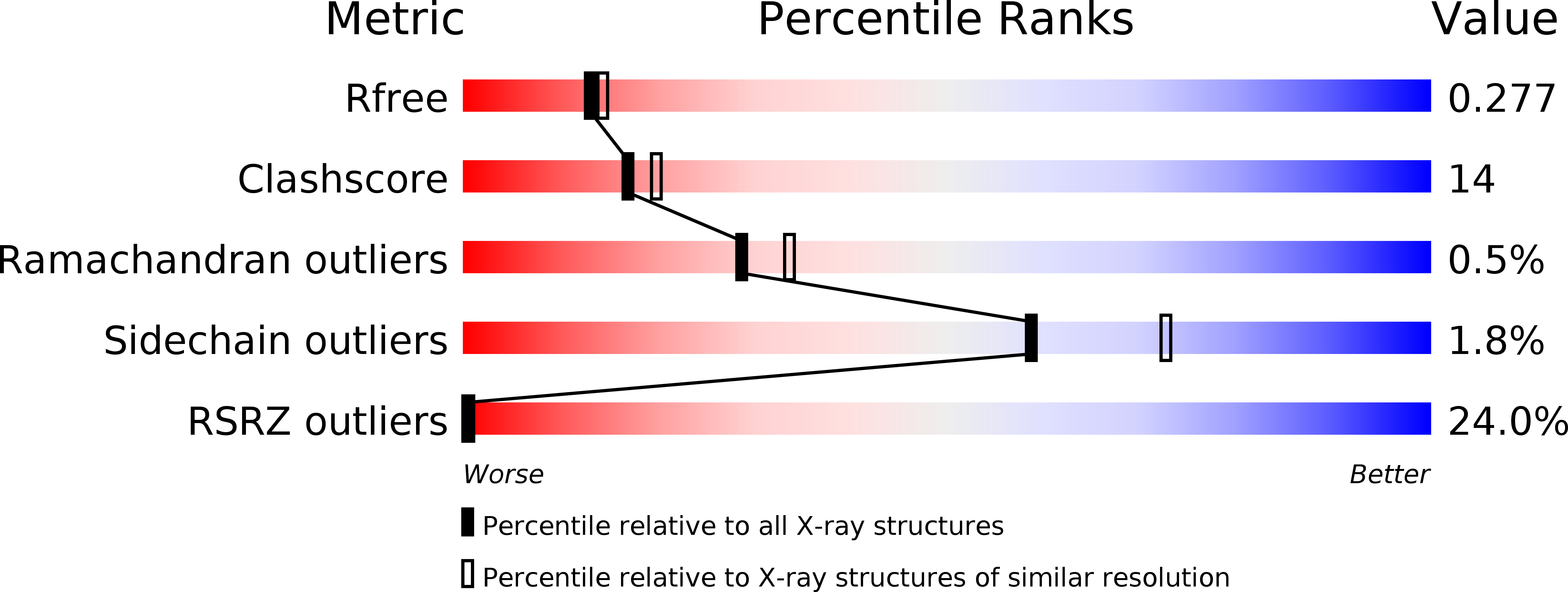
Resolution:
2.28 Å
R-Value Free:
0.27
R-Value Work:
0.24
Space Group:
P 32 2 1
Macromolecular Entities
Polymer Type:polypeptide(L)
Molecule:mRNA interferase toxin HicA
Gene (Uniprot):hicA
Chain IDs:A, C
Chain Length:58
Number of Molecules:2
Biological Source:Escherichia coli str. K-12 substr. MG1655
Polymer Type:polypeptide(L)
Molecule:Antitoxin HicB
Gene (Uniprot):hicB
Chain IDs:B, D
Chain Length:138
Number of Molecules:2
Biological Source:Escherichia coli 2-222-05_S4_C3
Modified Residue
Compound ID Chain ID Parent Comp ID Details 2D Image
MSE A MET modified residue
Ligand Molecules
Primary Citation
The E. coli HicB Antitoxin Contains a Structurally Stable Helix-Turn-Helix DNA Binding Domain.
Structure 27 1675 1685.e3 (2019)
PMID: 31495532 DOI: 10.1016/j.str.2019.08.008

Abstact

The E. coli hicAB type II toxin-antitoxin locus is unusual by being controlled by two promoters and by having the toxin encoded upstream of the antitoxin. HicA toxins contain a double-stranded RNA-binding fold and cleaves both mRNA and tmRNA in vivo, while HicB antitoxins contain a partial RNase H fold and either a helix-turn-helix (HTH) or ribbon-helix-helix domain. It is not known how an HTH DNA-binding domain affects higher-order structure for the HicAB modules. Here, we present crystal structures of the isolated E. coli HicB antitoxin and full-length HicAB complex showing that HicB forms a stable DNA-binding module and interacts in a canonical way with HicA despite the presence of an HTH-type DNA-binding domain. No major structural rearrangements take place upon binding of the toxin. Both structures expose well-ordered DNA-binding motifs allowing a model for DNA binding by the antitoxin to be generated.

Legend

Protein

Chemical

Disease

Primary Citation of related structures