6HMA image
Deposition Date 2018-09-12
Release Date 2018-11-14
Last Version Date 2025-10-22
Entry Detail
PDB ID:
6HMA
Keywords:
Title:
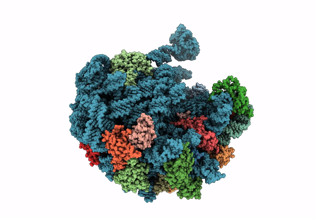
Improved model derived from cryo-EM map of Staphylococcus aureus large ribosomal subunit
Biological Source:
Source Organism:
Method Details:
Experimental Method:
Resolution:
2.65 Å
Aggregation State:
PARTICLE
Reconstruction Method:
SINGLE PARTICLE
Macromolecular Entities
Structures with similar UniProt ID
Protein Blast
Polymer Type:polypeptide(L)
Molecule:50S ribosomal protein L33
Chain IDs:Z (auth: 1)
Chain Length:47
Number of Molecules:1
Biological Source:Staphylococcus aureus
Structures with similar UniProt ID
Protein Blast
Polymer Type:polypeptide(L)
Molecule:50S ribosomal protein L34
Chain IDs:AA (auth: 2)
Chain Length:43
Number of Molecules:1
Biological Source:Staphylococcus aureus
Structures with similar UniProt ID
Protein Blast
Polymer Type:polypeptide(L)
Molecule:50S ribosomal protein L35
Chain IDs:BA (auth: 3)
Chain Length:64
Number of Molecules:1
Biological Source:Staphylococcus aureus
Structures with similar UniProt ID
Protein Blast
Polymer Type:polypeptide(L)
Molecule:50S ribosomal protein L36
Chain IDs:CA (auth: 4)
Chain Length:37
Number of Molecules:1
Biological Source:Staphylococcus aureus
Polymer Type:polyribonucleotide
Molecule:23S ribosomal RNA
Chain IDs:A
Chain Length:2923
Number of Molecules:1
Biological Source:Staphylococcus aureus
Polymer Type:polyribonucleotide
Molecule:5S ribosomal RNA
Chain IDs:B
Chain Length:115
Number of Molecules:1
Biological Source:Staphylococcus aureus
Structures with similar UniProt ID
Protein Blast
Polymer Type:polypeptide(L)
Molecule:50S ribosomal protein L2
Chain IDs:C
Chain Length:274
Number of Molecules:1
Biological Source:Staphylococcus aureus
Structures with similar UniProt ID
Protein Blast
Polymer Type:polypeptide(L)
Molecule:50S ribosomal protein L3
Chain IDs:D
Chain Length:215
Number of Molecules:1
Biological Source:Staphylococcus aureus
Structures with similar UniProt ID
Protein Blast
Polymer Type:polypeptide(L)
Molecule:50S ribosomal protein L4
Chain IDs:E
Chain Length:206
Number of Molecules:1
Biological Source:Staphylococcus aureus
Structures with similar UniProt ID
Protein Blast
Polymer Type:polypeptide(L)
Molecule:50S ribosomal protein L5
Chain IDs:F
Chain Length:158
Number of Molecules:1
Biological Source:Staphylococcus aureus
Structures with similar UniProt ID
Protein Blast
Polymer Type:polypeptide(L)
Molecule:50S ribosomal protein L6
Chain IDs:G
Chain Length:175
Number of Molecules:1
Biological Source:Staphylococcus aureus
Structures with similar UniProt ID
Protein Blast
Polymer Type:polypeptide(L)
Molecule:50S ribosomal protein L13
Chain IDs:H
Chain Length:145
Number of Molecules:1
Biological Source:Staphylococcus aureus
Structures with similar UniProt ID
Protein Blast
Polymer Type:polypeptide(L)
Molecule:50S ribosomal protein L14
Chain IDs:I
Chain Length:122
Number of Molecules:1
Biological Source:Staphylococcus aureus
Structures with similar UniProt ID
Protein Blast
Polymer Type:polypeptide(L)
Molecule:50S ribosomal protein L15
Chain IDs:J
Chain Length:146
Number of Molecules:1
Biological Source:Staphylococcus aureus
Structures with similar UniProt ID
Protein Blast
Polymer Type:polypeptide(L)
Molecule:50S ribosomal protein L16
Chain IDs:K
Chain Length:137
Number of Molecules:1
Biological Source:Staphylococcus aureus
Structures with similar UniProt ID
Protein Blast
Polymer Type:polypeptide(L)
Molecule:50S ribosomal protein L17
Chain IDs:L
Chain Length:120
Number of Molecules:1
Biological Source:Staphylococcus aureus
Structures with similar UniProt ID
Protein Blast
Polymer Type:polypeptide(L)
Molecule:50S ribosomal protein L18
Chain IDs:M
Chain Length:119
Number of Molecules:1
Biological Source:Staphylococcus aureus
Structures with similar UniProt ID
Protein Blast
Polymer Type:polypeptide(L)
Molecule:50S ribosomal protein L19
Chain IDs:N
Chain Length:116
Number of Molecules:1
Biological Source:Staphylococcus aureus
Structures with similar UniProt ID
Protein Blast
Polymer Type:polypeptide(L)
Molecule:50S ribosomal protein L20
Chain IDs:O
Chain Length:116
Number of Molecules:1
Biological Source:Staphylococcus aureus
Structures with similar UniProt ID
Protein Blast
Polymer Type:polypeptide(L)
Molecule:50S ribosomal protein L21
Chain IDs:P
Chain Length:102
Number of Molecules:1
Biological Source:Staphylococcus aureus
Structures with similar UniProt ID
Protein Blast
Polymer Type:polypeptide(L)
Molecule:50S ribosomal protein L22
Chain IDs:Q
Chain Length:112
Number of Molecules:1
Biological Source:Staphylococcus aureus
Structures with similar UniProt ID
Protein Blast
Polymer Type:polypeptide(L)
Molecule:50S ribosomal protein L23
Chain IDs:R
Chain Length:89
Number of Molecules:1
Biological Source:Staphylococcus aureus
Structures with similar UniProt ID
Protein Blast
Polymer Type:polypeptide(L)
Molecule:50S ribosomal protein L24
Chain IDs:S
Chain Length:103
Number of Molecules:1
Biological Source:Staphylococcus aureus
Structures with similar UniProt ID
Protein Blast
Polymer Type:polypeptide(L)
Molecule:50S ribosomal protein L25
Chain IDs:T
Chain Length:94
Number of Molecules:1
Biological Source:Staphylococcus aureus
Structures with similar UniProt ID
Protein Blast
Polymer Type:polypeptide(L)
Molecule:50S ribosomal protein L27
Chain IDs:U
Chain Length:79
Number of Molecules:1
Biological Source:Staphylococcus aureus
Structures with similar UniProt ID
Protein Blast
Polymer Type:polypeptide(L)
Molecule:50S ribosomal protein L28
Chain IDs:V
Chain Length:49
Number of Molecules:1
Biological Source:Staphylococcus aureus
Structures with similar UniProt ID
Protein Blast
Polymer Type:polypeptide(L)
Molecule:50S ribosomal protein L29
Chain IDs:W
Chain Length:67
Number of Molecules:1
Biological Source:Staphylococcus aureus
Structures with similar UniProt ID
Protein Blast
Polymer Type:polypeptide(L)
Molecule:50S ribosomal protein L30
Chain IDs:X
Chain Length:58
Number of Molecules:1
Biological Source:Staphylococcus aureus
Structures with similar UniProt ID
Protein Blast
Polymer Type:polypeptide(L)
Molecule:50S ribosomal protein L32
Chain IDs:Y (auth: Z)
Chain Length:48
Number of Molecules:1
Biological Source:Staphylococcus aureus
Modified Residue
Compound ID Chain ID Parent Comp ID Details 2D Image
2MA A A modified residue
5MU A U modified residue
Ligand Molecules
Primary Citation
Structural Studies Reveal the Role of Helix 68 in the Elongation Step of Protein Biosynthesis.
Mbio 13 e0030622 e0030622 (2022)
PMID: 35348349 DOI: 10.1128/mbio.00306-22

Abstact

The ribosome, a multicomponent assembly consisting of RNA and proteins, is a pivotal macromolecular machine that translates the genetic code into proteins. The large ribosomal subunit rRNA helix 68 (H68) is a key element in the protein synthesis process, as it coordinates the coupled movements of the actors involved in translocation, including the tRNAs and L1 stalk. Examination of cryo-electron microscopy (cryo-EM) structures of ribosomes incubated for various time durations at physiological temperatures led to the identification of functionally relevant H68 movements. These movements assist the transition of the L1 stalk between its open and closed states. H68 spatial flexibility and its significance to the protein synthesis process were confirmed through its effective targeting with antisense PNA oligomers. Our results suggest that H68 is actively involved in ribosome movements that are central to the elongation process. IMPORTANCE The mechanism that regulates the translocation step in ribosomes during protein synthesis is not fully understood. In this work, cryo-EM techniques used to image ribosomes from Staphylococcus aureus after incubation at physiological temperature allowed the identification of a conformation of the helix 68 that has never been observed so far. We then propose a mechanism in which such helix, switching between two different conformations, actively coordinates the translocation step, shedding light on the dynamics of ribosomal components. In addition, the relevance of helix 68 to ribosome function and its potential as an antibiotic target was proved by inhibiting Staphylococcus aureus ribosomes activity in vitro using oligomers with sequence complementarity.

Legend

Protein

Chemical

Disease

Primary Citation of related structures